is a curved glass that bends light that passes through it. regulates the amount of light on the specimen. The glass slide is placed on the for viewing.
is a curved glass that bends light that passes through it. regulates the amount of light on the specimen. The glass slide is placed on the for viewing.

Fundamentals of Sectional Anatomy: An Imaging Approach
2nd Edition
ISBN:9781133960867
Author:Denise L. Lazo
Publisher:Denise L. Lazo
Chapter3: Face
Section: Chapter Questions
Problem 10RQ

Related questions
Question
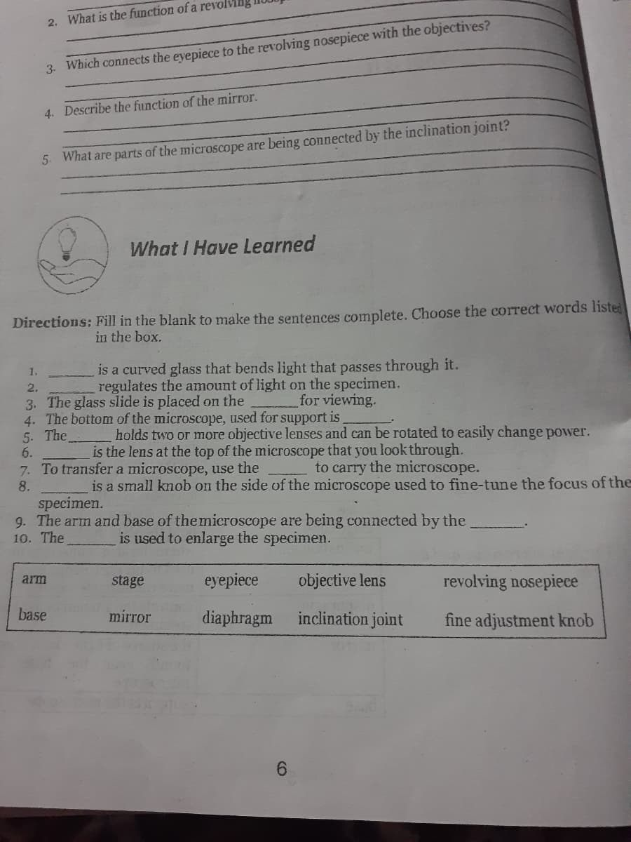
Transcribed Image Text:2. What is the function of a revolvillg
3. Which connects the eyepiece to the revolving nosepiece with the objectives?
4. Describe the function of the mirror.
5 What are parts of the microscope are being connected by the inclination joint?
What I Have Learned
Directions: Fill in the blank to make the sentences complete. Choose the correct words listed
in the box.
is a curved glass that bends light that passes through it.
regulates the amount of light on the specimen.
1,
2.
for viewing.
3. The glass slide is placed on the
4. The bottom of the microscope, used for support is
5. The
6.
7. To transfer a microscope, use the
holds two or more objective lenses and can be rotated to easily change power.
is the lens at the top of the microscope that you look through.
to carry the microscope.
8.
is a small knob on the side of the microscope used to fine-tune the focus of the
specimen.
9. The arm and base of themicroscope are being connected by the
10. The
is used to enlarge the specimen.
stage
eyepiece
objective lens
revolving nosepiece
arm
base
mirror
diaphragm
inclination joint
fine adjustment knob
Expert Solution
This question has been solved!
Explore an expertly crafted, step-by-step solution for a thorough understanding of key concepts.

This is a popular solution!

Trending now
This is a popular solution!

Step by step
Solved in 2 steps

Recommended textbooks for you
Fundamentals of Sectional Anatomy: An Imaging App…
Biology
ISBN:
9781133960867
Author:
Denise L. Lazo
Publisher:
Cengage Learning
Fundamentals of Sectional Anatomy: An Imaging App…
Biology
ISBN:
9781133960867
Author:
Denise L. Lazo
Publisher:
Cengage Learning