
Videos
Connecting the Concepts
1. Use the following diagram to review the flow of blood through a human cardiovascular system. Label the indicated parts, highlight the vessels that carry oxygen-rich blood, and then trace the flow of blood by numbering the circles from 1 to 10, starting with 1 in the right ventricle. (When two locations are equivalent in the pathway, such as right and left lung capillaries or capillaries of top and lower portion of the body, assign them the same number.)
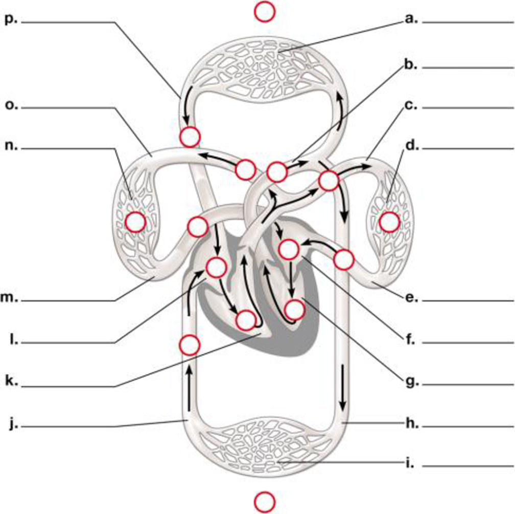
To label: The diagram of the flow of blood through the circulatory system.
Concept introduction:
The blood flows through the circulatorysystem. It includes various capillaries, aorta, pulmonary artery, pulmonary veins, ventricles, auricles, superior and inferior vena cava.
Answer to Problem 1CC
Pictorial representation: A labelled diagram of human circulatory system showing the flow of blood through it is given on Fig.1.

Fig. 1: The circulatory system.
Explanation of Solution
The labeling of the blood vessels in the circulatory system include as follows:
(a)
Network of the blood capillaries: It supplies blood to the various parts of the human body like head, arms, chest and legs.
(b)
Aorta: The blood moves from the heart chambers through this blood vessel.
(c)
Pulmonary artery: It carries the deoxygenated blood.
(d)
Capillaries: They supply blood to the lungs: The blood flows through them and reaches the lungs.
(e)
Pulmonary vein: It carries oxygenated blood.
(f)
Left atrium: It is the upper chamber of the heart which contracts to cause the flow of the blood out of the heart.
(g)
Left ventricle: It is the lower chamber of the heart where the blood flows.
(h)
Aorta: This blood vessel transports the blood to the organs from the heart.
(i)
Capillaries: They supply the blood of abdominal regions and legs:
(j)
Inferior vena cava: It drains the blood into the right atrium.
(k)
Right ventricle: It is the lower chamber of the heart.
(l)
Right atrium: It is the upper chamber of the heart.
(m)
Pulmonary vein: The left atrium of the heart receives blood from the pulmonary vein.
(n)
Capillaries of right lung: It helps in the flow of the blood to the lungs.
(o)
Pulmonary artery: It carries deoxygenated blood.
(p)
Superior vena cava: It circulates the blood in the sino-atrial node in the right atrium.
Want to see more full solutions like this?
Chapter 23 Solutions
CAMPBELL BIOLOGY - BIO 121 W/ACC >IC<


- Figure 40.11 Which of the following statements about the heart is false? The mitral valve separates the left ventricle from the left atrium. Blood travels through the bicuspid valve to the left atrium. Both the aortic and the pulmonary valves are semilunar valves. The mitral valve is an atrioventricular valve.arrow_forward5. Singh then created a flow chart of the blood returning to the right atrium by the vena cava to being ejected into the aorta. Using the diagram on the next page, fill in the missing information as Singh would have using the word bank below: Aorta Pulmonary veins Right ventricle Pulmonary semilunar valves Word Bank Coronary arteries Pulmonary arteries Left ventricle Bicuspid valve "The Breathless Heart" by Broadbelt, Sawhney, & Young Superior and Inferior venae Right Atrium Tricuspid valve cavae REST OF THE BODY HEART Coronary veins and sinus Pulmonary trunk Left atrium Aortic semilunar valves Page 2 NATIONAL CENTER FOR CASE STUDY TEACHING IN SCIENCE LUNGarrow_forwardConceptualize in Color Principal Arteries Color these principal arteries in the following figure using the colors suggested, or choose your own. • Subclavian: Red • Axillary: Green • Brachial: Orange • Radial: Purple • Celiac trunk: Red • Superior mesenteric: Blue • Inferior mesenteric: Yellow • Renal: Brown • Common iliae: Pink • Internal iliac: Green • External iliac: Orange • Femoral: Purple • Popliteal: Green • Anterior tibial: Blue • Posterior tibial: Orange • Dorsalis pedis: Red 204 Chapter 16 Vascular Systemarrow_forward
- Conceptualize in Color The Aorta Color the regions of the aorta and its branches susing the colors suggested, or choose your own. • Ascending aorta: Blue • Aortic arch: Green • Descending aorta: Pink • Abdominal aorta: Red • Right common carotid: Orange • Left common carotid: Orange • Left subclavian artery: Purple • Right subclavian artery: Purple • Brachiocephalic artery: Brown • Right and left common iliac arteries: Yellow Chapter 16 Vascular System 203arrow_forward5. Describe the conductions of electrical signals through the heart. answer this question with a narrative of the structures involved in initiating and coordinating the contraction of the heart. You need to include the function of each structure. Mention pacemaker cells, the sinoatrial node, atrioventricular node, internodal pathway, bundle of His, bundle branches, Purkinje fibers, first degree block, second degree block, and third degree block.arrow_forward2. Make a table listing all the parts and functions of the Circulatory system. the 3. Describe the process done during Pulmonary and Systemic Circulation. Use illustration provided below. Source: Human Physiology from Cells to Systems seventh Capilary bed of lungs where gas exchange occurs Pulmonary arteries Pulmonary veins Pulmonary circuit Aorta and branches Vena cavae Left atrium Left ventricle Right atrium Right ventricle Systemic arteries Systemic veins Охудел роог, CO, - rich błood Oxygen rich, CO, - poor blood Systemic circuit Capilary bed of all body tissues where Lesson 1.: item gas exchange Occurs |Pagearrow_forward
- 40- In the brain the frontal lobe has Oa. Voluntary motor activity Ob. Speaking Oc. Reasoning Od. Problem solving Oe. All of the abovearrow_forwardLet's Review - Human Circulation Your textbook begins the discussion of the path that blood takes around the body starting at the right atrium. But remember that this path a cycle, and therefore there is no true beginning or end. To make sure you have the idea, I would like you to trace the flow of blood around the human body starting at the AORTA. Complete the activity below. 1 E Right Atrium E Right ventricle 3 Aorta 4 Left Ventricle 5 : Left Atrium 6 Vena Cava 7 | Cells of the body 8 | Lungs ::::arrow_forwardPut the following in proper order of blood flow for pulmonary and systemic circulation. 1 [ Choose ] [Choose ] Capillaries at the systemic organs (e.g. liver or kidney) Left side of the heart Right side of the heart Lungs 3 [ Choose ] 4 [ Choose ]arrow_forward
- Select TRUE or FALSE to best describe the statements below, relating the events of the cardiac cycle. Keep in mind that these events may be chemical, electrical and mechanical. atrial pressure is greater than ventricular pressure while semilunar valves are оpen ventricular pressure is greater than atrial pressure when the atrioventricular valves are closed atrioventricular valves are open during ventricular emptying the end diastolic volume of the ventricles is reached before ventricles contract 1 ventricular filling occurs before depolarization of the atria 1 atrioventricular valves and 1. TRUE 1 semilunar valves are closed at the beginning of ventricular systole 2. FALSE end systolic volume increases when more blood empties from the ventricles the greatest pressure is reached in the ventricles while they are emptying the right atrium depolarizes before the left atrium ventricular depolarization occurs during the T-wave semilunar valves are open during ventricular filling ventricular…arrow_forwarddescribe and explain 2 physical ways that a human may develop hypertension (abnormally elevated blood pressure). [One of these mechanisms should stem from the blood vessels, and the other at the level of the nervous system]. What are the long-term consequences of elevated hypertension?arrow_forwardIn the given table, three of the anatomical and physiological terms are similar or related; one does not belong with the other three. Choose the term that does NOT belong in each of the following groups. A B C D 1 Pulmonary Trunk Vena Cava Right Side of the Heart Left Side of the Heart 2 QRS Wave T Wave P Wave Electrical Activity of the Ventricles 3 AV Valves Closed AV Valves Opened Ventricular Systole Semilunar Valves Open 4 Tricuspid Valve Mitral Valve Bicuspid Valve Left AV Valve 5 Pulmonary Valve Umbilical Artery Pulmonary Vein Superior Vena Cavaarrow_forward
 Biology 2eBiologyISBN:9781947172517Author:Matthew Douglas, Jung Choi, Mary Ann ClarkPublisher:OpenStax
 Human Physiology: From Cells to Systems (MindTap ...BiologyISBN:9781285866932Author:Lauralee SherwoodPublisher:Cengage Learning