1. What kinds of media would be used to culture and Identify this microbe? 2. What are some other potential microbes that could have this infection?
1. What kinds of media would be used to culture and Identify this microbe? 2. What are some other potential microbes that could have this infection?

Surgical Tech For Surgical Tech Pos Care
5th Edition
ISBN:9781337648868
Author:Association
Publisher:Association
Chapter7: Preventing Perioperative Disease Transmission
Section: Chapter Questions
Problem 3CS

Related questions

Question
100%
1. What kinds of media would be used to culture and Identify this microbe?
2. What are some other potential microbes that could have this infection?
Transcribed Image Text:CASE STUDY Part 2
Micro
riety
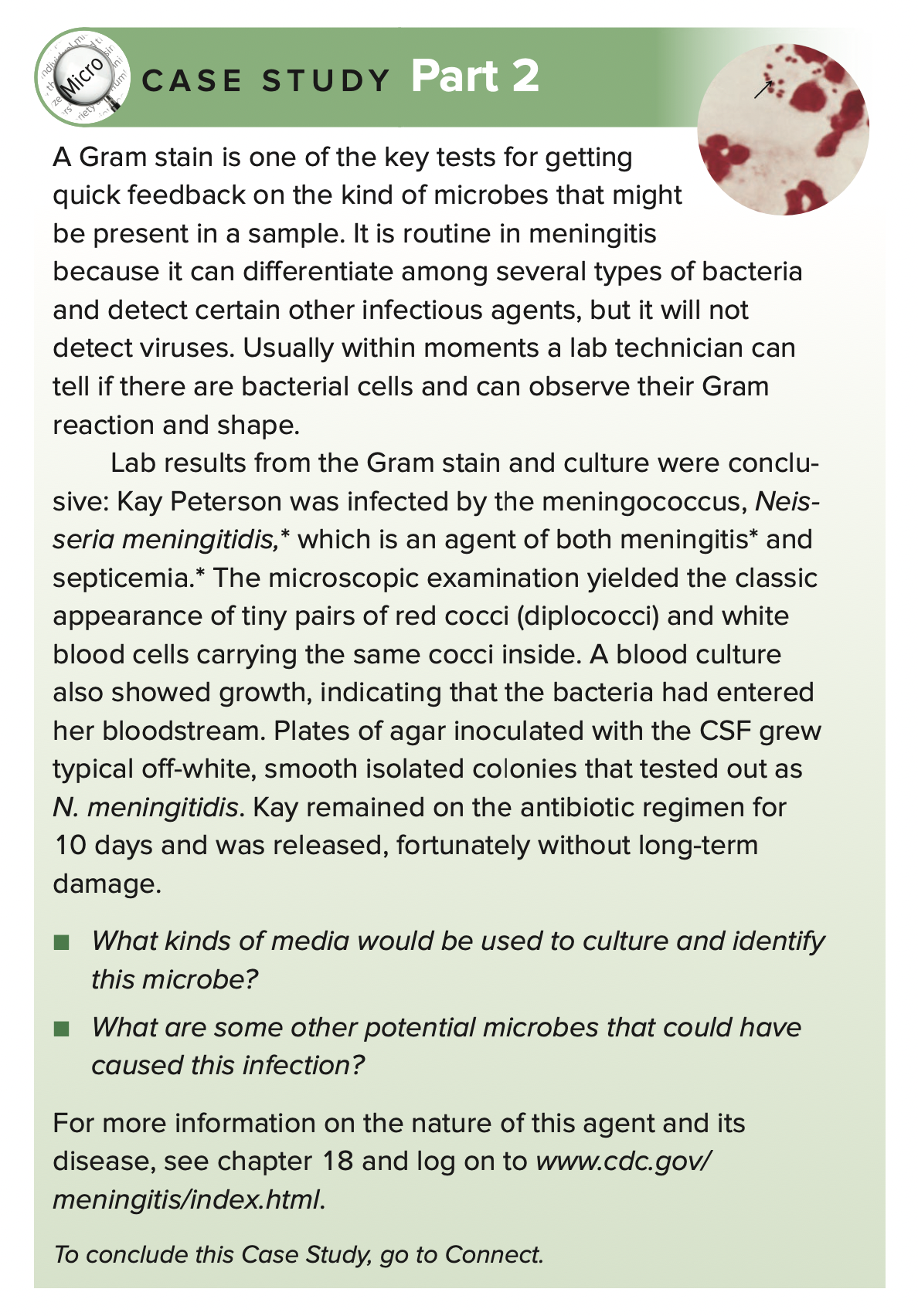
A Gram stain is one of the key tests for getting
quick feedback on the kind of microbes that might
be present in a sample. It is routine in meningitis
because it can differentiate among several types of bacteria
and detect certain other infectious agents, but it will not
detect viruses. Usually within moments a lab technician can
tell if there are bacterial cells and can observe their Gram
reaction and shape.
Lab results from the Gram stain and culture were conclu-
sive: Kay Peterson was infected by the meningococcus, Neis-
seria meningitidis,* which is an agent of both meningitis* and
septicemia.* The microscopic examination yielded the classic
appearance of tiny pairs of red cocci (diplococci) and white
blood cells carrying the same cocci inside. A blood culture
also showed growth, indicating that the bacteria had entered
her bloodstream. Plates of agar inoculated with the CSF grew
typical off-white, smooth isolated colonies that tested out as
N. meningitidis. Kay remained on the antibiotic regimen for
10 days and was released, fortunately without long-term
damage.
What kinds of media would be used to culture and identify
this microbe?
What are some other potential microbes that could have
caused this infection?
For more information on the nature of this agent and its
disease, see chapter 18 and log on to www.cdc.gov/
meningitis/index.html.
To conclude this Case Study, go to Connect.
ndivi
Expert Solution
This question has been solved!
Explore an expertly crafted, step-by-step solution for a thorough understanding of key concepts.

This is a popular solution!

Trending now
This is a popular solution!

Step by step
Solved in 2 steps

Knowledge Booster


Learn more about
Need a deep-dive on the concept behind this application? Look no further. Learn more about this topic, biology and related others by exploring similar questions and additional content below.Recommended textbooks for you
Surgical Tech For Surgical Tech Pos Care
Health & Nutrition
ISBN:
9781337648868
Author:
Association
Publisher:
Cengage
Surgical Tech For Surgical Tech Pos Care
Health & Nutrition
ISBN:
9781337648868
Author:
Association
Publisher:
Cengage