compare that hypothetical mechanism to the classical presentation described in our textbook. What are the major differences between this mechanism and Peter Mitchel’s original chemiosmotic theory? What are the similarities.
compare that hypothetical mechanism to the classical presentation described in our textbook. What are the major differences between this mechanism and Peter Mitchel’s original chemiosmotic theory? What are the similarities.

Biology: The Unity and Diversity of Life (MindTap Course List)
15th Edition
ISBN:9781337408332
Author:Cecie Starr, Ralph Taggart, Christine Evers, Lisa Starr
Publisher:Cecie Starr, Ralph Taggart, Christine Evers, Lisa Starr
Chapter7: Releasing Chemical Energy
Section: Chapter Questions
Problem 10SQ: Which of the following is not produced by an animal muscle cell operating under anaerobic...

Related questions
Question
Focusing on the mechanism linking complex I and ATP synthase depicted in figure 3 in the article, compare that hypothetical mechanism to the classical presentation described in our textbook. What are the major differences between this mechanism and Peter Mitchel’s original chemiosmotic theory? What are the similarities.
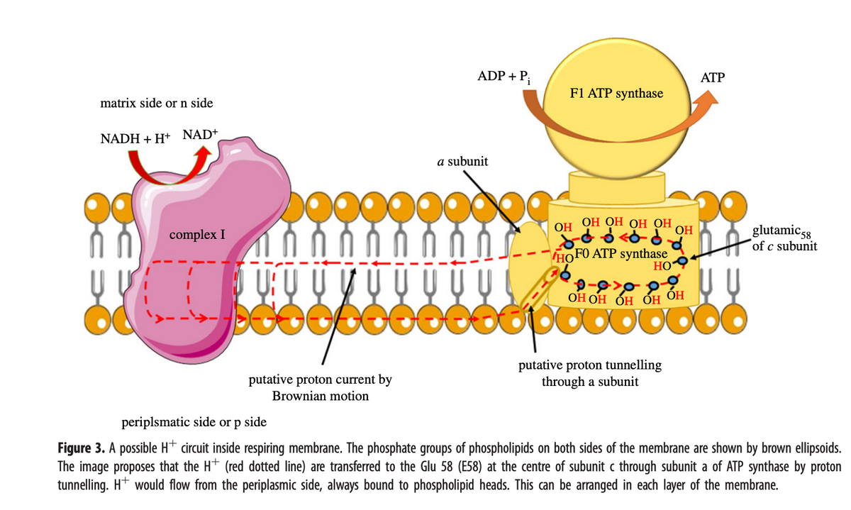
Transcribed Image Text:matrix side or n side
NADH+H+ NAD+
complex I
VUU
ooo
putative proton current by
Brownian motion
ADP + Pi
a subunit
DG
F1 ATP synthase
ОН ОН ОН ОН
<d
HOFO ATP synthase
HO
OH
OH
putative proton tunnelling
through a subunit
ATP
U
ОНОН ОН ОН ОН
beepooooooo
glutamic 58
of c subunit
periplsmatic side or p side
Figure 3. A possible H* circuit inside respiring membrane. The phosphate groups of phospholipids on both sides of the membrane are shown by brown ellipsoids.
The image proposes that the H+ (red dotted line) are transferred to the Glu 58 (E58) at the centre of subunit c through subunit a of ATP synthase by proton
tunnelling. H+ would flow from the periplasmic side, always bound to phospholipid heads. This can be arranged in each layer of the membrane.
Transcribed Image Text:+
b.
+
Inner
membrane
+
+
4 H
+ +
4 H+
2 e
NAD+
Inner membrane
+ + +
+
Dihydroxyacetone phosphate
Glycerol-3-P
dehydrogenase
Succinate
dehydrogenase
+
2 e
NADH + H+
-2 H+
FADH,
Citrate
cycle
+
Fumarate Succinate
QH₂
NADH-ubiquinone
oxidoreductase
FADH₂
Glycerol-3-P
2 e
+
2 e
+
FADH₂
2 H
Acetyl-CoA
+
QH₂
Fatty
acids
2 H
+
2 e
4 H
Ubiquinone-cytochrome c
oxidoreductase
2 H
2 x 1 e
4 H
ETF-Q
oxidoreductase
Cyt c
2 x 1e Cyt c
+ +
+
Intermembrane
space
2 x 1 e
2 e
2 H+ + O₂ H₂O
IV
2 H+
Cytochrome c oxidase
Mitochondrial
matrix
Intermembrane
space
2x1 e
IV
2 e
2 H+
2 H+ O₂ H₂O
matrix
2 H
2H+
Mitochondrial
+
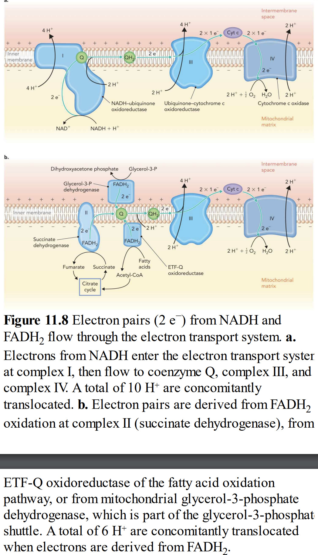
Figure 11.8 Electron pairs (2 e) from NADH and
FADH₂ flow through the electron transport system. a.
Electrons from NADH enter the electron transport syster
at complex I, then flow to coenzyme Q, complex III, and
complex IV. A total of 10 H* are concomitantly
translocated. b. Electron pairs are derived from FADH₂
oxidation at complex II (succinate dehydrogenase), from
ETF-Q oxidoreductase of the fatty acid oxidation
pathway, or from mitochondrial glycerol-3-phosphate
dehydrogenase, which is part of the glycerol-3-phosphat
shuttle. A total of 6 H* are concomitantly translocated
when electrons are derived from FADH₂.
Expert Solution
This question has been solved!
Explore an expertly crafted, step-by-step solution for a thorough understanding of key concepts.

Step by step
Solved in 4 steps

Recommended textbooks for you
Biology: The Unity and Diversity of Life (MindTap…
Biology
ISBN:
9781337408332
Author:
Cecie Starr, Ralph Taggart, Christine Evers, Lisa Starr
Publisher:
Cengage Learning

Biology 2e
Biology
ISBN:
9781947172517
Author:
Matthew Douglas, Jung Choi, Mary Ann Clark
Publisher:
OpenStax

Biochemistry
Biochemistry
ISBN:
9781305577206
Author:
Reginald H. Garrett, Charles M. Grisham
Publisher:
Cengage Learning
Biology: The Unity and Diversity of Life (MindTap…
Biology
ISBN:
9781337408332
Author:
Cecie Starr, Ralph Taggart, Christine Evers, Lisa Starr
Publisher:
Cengage Learning
Biology 2e
Biology
ISBN:
9781947172517
Author:
Matthew Douglas, Jung Choi, Mary Ann Clark
Publisher:
OpenStax
Biochemistry
Biochemistry
ISBN:
9781305577206
Author:
Reginald H. Garrett, Charles M. Grisham
Publisher:
Cengage Learning