During an MRI, when the radio signal is removed and the hydrogen protons re-align with the magnetic field, they emit their own radio waves. Suggest why this might occur.
During an MRI, when the radio signal is removed and the hydrogen protons re-align with the magnetic field, they emit their own radio waves. Suggest why this might occur.

Organic Chemistry
8th Edition
ISBN:9781305580350
Author:William H. Brown, Brent L. Iverson, Eric Anslyn, Christopher S. Foote
Publisher:William H. Brown, Brent L. Iverson, Eric Anslyn, Christopher S. Foote
Chapter12: Infrared Spectroscopy
Section12.3: Infrared Spectroscopy
Problem 12.1P

Related questions
Question
Answer in a detailed paragraph

Transcribed Image Text:2. During an MRI, when the radio signal is removed and the hydrogen
protons re-align with the magnetic field, they emit their own radio
waves. Suggest why this might occur.
Transcribed Image Text:Exit Pullscreen
Enhanced MRI Images and Epilepsy Research
Four to five of every one thousand Canadians
have epilepsy, a disorder characterized by
sudden changes in brain function that often
result in seizures. Thirty percent of individuals
with epilepsy do not respond to medications
and are diagnosed with medically-intractable
epilepsy. Dr. Jorge Burneo of the University
of Western Ontario in London, Ontario
hopes to better understand the causes of
this condition with the help of the newest
addition to the university's research arsenal,
a 7 Tesla MRI. MRI (magnetic resonance
imaging) technology uses radio waves along
with a powerful magnet and a computer to
generate images of soft tissue. Like electrons,
protons spin on their axes. However, the
orientation of these axes is random. The
strong magnetic field generated by the MRI
magnet affects the protons in hydrogen
atoms in body tissue, causing their axes to
align with the same orientation. This is similar
to how the needles in compasses adopt a
north-south alignment when exposed to
Earth's magnetic field. When exposed to
radio waves at a specific frequency, certain
protons are momentarily "knocked" out of this
alignment. When the radio signal is removed,
these protons re-align with the magnetic field,
emitting their own radio waves as they do so.
These signals are "read" by the computer to
produce a detailed image of the body tissue.
Image resolution depends on the strength of
the magnet, which is measured in teslas, the
SI unit of measurement for the concentration
of a magnetic field. Used only for research
purposes, the 7 Telsa is the world's most
powerful MRI and the only one of its kind in
Canada.
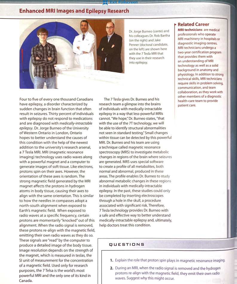
Dr. Jorge Burneo (cente) and
his colleagues Dr. Rob Bartha
(on the right) and Jake
Penner (doctoral candidate,
on the left) are shown here
with the 7 Tesla MRI that
they use in their research
into epilepsy.
The 7 Tesla gives Dr. Burneo and his
research team a glimpse into the brains
of individuals with medically-intractable
epilepsy in a way that less-powerful MRIs
cannot. "We hope," Dr. Burneo states, "that
with the use of the 7T technology, we will
be able to identify structural abnormalities
not seen in standard testing." Small changes
within tissue can be detected by this powerful
MRI. Dr. Burneo and his team are using
a technique called magnetic resonance
spectroscopy (MRS) to investigate metabolic
changes in regions of the brain where seizures
are generated. MRS uses special software
to create a profile of all metabolites, both
normal and abnormal, produced in these
areas. The profile enables Dr. Burneo to study
abnormal metabolic changes in these regions
in individuals with medically-intractable
epilepsy. In the past, these studies could only
be completed by inserting electroscopes
through a hole in the skull, a procedure
associated with significant risk. Therefore,
7 Tesla technology provides Dr. Burneo with
a safe and effective way to better understand
medically-intractable epilepsy and, ultimately,
help doctors treat this condition.
QUESTIONS
Related Career
MRI technicians are medical
professionals who operate
MRI machinery in hospitals or
diagnostic imaging centres.
MRI technicians undergo a
two-year certification program
that provides them with
an understanding of MRI
technology as well as a solid
background in anatomy and
physiology. In addition to strong
technical skills, MRI technicians
require skills in problem solving,
communication, and team
collaboration, as they work with
other members of a diagnostic
health-care team to provide
patient care.
1. Explain the role that proton spin plays in magnetic resonance imaging.
2. During an MRI, when the radio signal is removed and the hydrogen
protons re-align with the magnetic field, they emit their own radio
waves. Suggest why this might occur.
Expert Solution
This question has been solved!
Explore an expertly crafted, step-by-step solution for a thorough understanding of key concepts.

Step by step
Solved in 3 steps

Knowledge Booster


Learn more about
Need a deep-dive on the concept behind this application? Look no further. Learn more about this topic, chemistry and related others by exploring similar questions and additional content below.Recommended textbooks for you
Organic Chemistry
Chemistry
ISBN:
9781305580350
Author:
William H. Brown, Brent L. Iverson, Eric Anslyn, Christopher S. Foote
Publisher:
Cengage Learning

Principles of Instrumental Analysis
Chemistry
ISBN:
9781305577213
Author:
Douglas A. Skoog, F. James Holler, Stanley R. Crouch
Publisher:
Cengage Learning
Organic Chemistry
Chemistry
ISBN:
9781305580350
Author:
William H. Brown, Brent L. Iverson, Eric Anslyn, Christopher S. Foote
Publisher:
Cengage Learning
Principles of Instrumental Analysis
Chemistry
ISBN:
9781305577213
Author:
Douglas A. Skoog, F. James Holler, Stanley R. Crouch
Publisher:
Cengage Learning