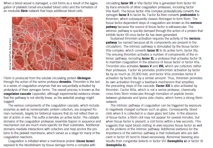
When a blood vessel is damaged, a clot forms as a result of the aggre- gation of platelets (small enucleated blood cells) and the formation of an insoluble fibrin network that traps additional blood cells. circulating factor VIl or Vla (factor Vla is generated from factor VII by trace amounts of other coagulation proteases, including factor VIlla itself). The tissue factor-VIla complex proteolytically converts the zymogen factor X to factor Xa. Factor Xa then converts prothrombin to thrombin, which subsequently cleaves fibrinogen to form fibrin. The tissue factor-dependent steps of coagulation are known as the extrinsic pathway because the source of tissue factor is extravascular. The extrinsic pathway is quickly damped through the action of a protein that inhibits factor VII once factor Xa has been generated. Sustained thrombin activation requires the activity of the intrinsic pathway (so named because all its components are present in the circulation). The intrinsic pathway is stimulated by the tissue factor- Vlla complex, which converts factor IX to its active form, factor IXa. The ensuing thrombin activates a number of components of the in- trinsic pathway, including factor XI, a protease that activates factor IX, to maintain coagulation in the absence of tissue factor or factor Vlla. Thrombin also activates factors V and VIII, which are cofactors rather than proteases. Factor Va promotes prothrombin activation by factor Xa by as much as 20,000-fold, and factor VIlla promotes factor X activation by factor IXa by a similar amount. Thus, thrombin promotes its own activation through a feedback mechanism that amplifies the preceding steps of the cascade. Factor XIII is also activated by thrombin. Factor XIlla, which is not a serine protease, chemically cross-links fibrin molecules through formation of peptide bonds between glutamate and lysine side chains, which forms a strong fibrin Fibrin is produced from the soluble circulating protein fibrinogen through the action of the serine protease thrombin. Thrombin is the last in a series of coagulation enzymes that are sequentially activated by proteolysis of their zymogen forms. The overall process is known as the coagulation cascade (opposite), although experimental evidence shows that the pathway is not strictly linear, as the waterfall analogy might network. The intrinsic pathway of coagulation can be triggered by exposure to negatively charged surfaces such as glass. Consequently, blood clots when it is collected in a clean glass test tube. In the absence of tissue factor, a fibrin clot may not appear for several minutes, but The various components of the coagulation cascade, which include enzymes as well as nonenzymatic protein cofactors, are assigned Ro- man numerals, largely for historical reasons that do not reflect their or- der of action in vivo. The suffix a denotes an active factor. The catalytic domains of the coagulation proteases resemble trypsin in sequence and when tissue factor is present, a clot forms within a few seconds. This mechanism but are much more specific for their substrates. Additional domains mediate interactions with cofactors and help anchor the pro- teins to the platelet membrane, which serves as a stage for many of the coagulation reactions. Coagulation is initiated when a membrane protein (tissue factor) exposed to the bloodstream by tissue damage forms a complex with suggests that rapid blood clotting in vivo requires tissue factor as well as the proteins of the intrinsic pathway. Additional evidence for the importance of the extrinsic pathway is that individuals who are defi- cient in factor VII tend to bleed excessively. Abnormal bleeding also results from congenital defects in factor VIII (hemophilia a) or factor IX (hemophilia b). Andrew Syred/Photo Resear chers
When a blood vessel is damaged, a clot forms as a result of the aggre- gation of platelets (small enucleated blood cells) and the formation of an insoluble fibrin network that traps additional blood cells. circulating factor VIl or Vla (factor Vla is generated from factor VII by trace amounts of other coagulation proteases, including factor VIlla itself). The tissue factor-VIla complex proteolytically converts the zymogen factor X to factor Xa. Factor Xa then converts prothrombin to thrombin, which subsequently cleaves fibrinogen to form fibrin. The tissue factor-dependent steps of coagulation are known as the extrinsic pathway because the source of tissue factor is extravascular. The extrinsic pathway is quickly damped through the action of a protein that inhibits factor VII once factor Xa has been generated. Sustained thrombin activation requires the activity of the intrinsic pathway (so named because all its components are present in the circulation). The intrinsic pathway is stimulated by the tissue factor- Vlla complex, which converts factor IX to its active form, factor IXa. The ensuing thrombin activates a number of components of the in- trinsic pathway, including factor XI, a protease that activates factor IX, to maintain coagulation in the absence of tissue factor or factor Vlla. Thrombin also activates factors V and VIII, which are cofactors rather than proteases. Factor Va promotes prothrombin activation by factor Xa by as much as 20,000-fold, and factor VIlla promotes factor X activation by factor IXa by a similar amount. Thus, thrombin promotes its own activation through a feedback mechanism that amplifies the preceding steps of the cascade. Factor XIII is also activated by thrombin. Factor XIlla, which is not a serine protease, chemically cross-links fibrin molecules through formation of peptide bonds between glutamate and lysine side chains, which forms a strong fibrin Fibrin is produced from the soluble circulating protein fibrinogen through the action of the serine protease thrombin. Thrombin is the last in a series of coagulation enzymes that are sequentially activated by proteolysis of their zymogen forms. The overall process is known as the coagulation cascade (opposite), although experimental evidence shows that the pathway is not strictly linear, as the waterfall analogy might network. The intrinsic pathway of coagulation can be triggered by exposure to negatively charged surfaces such as glass. Consequently, blood clots when it is collected in a clean glass test tube. In the absence of tissue factor, a fibrin clot may not appear for several minutes, but The various components of the coagulation cascade, which include enzymes as well as nonenzymatic protein cofactors, are assigned Ro- man numerals, largely for historical reasons that do not reflect their or- der of action in vivo. The suffix a denotes an active factor. The catalytic domains of the coagulation proteases resemble trypsin in sequence and when tissue factor is present, a clot forms within a few seconds. This mechanism but are much more specific for their substrates. Additional domains mediate interactions with cofactors and help anchor the pro- teins to the platelet membrane, which serves as a stage for many of the coagulation reactions. Coagulation is initiated when a membrane protein (tissue factor) exposed to the bloodstream by tissue damage forms a complex with suggests that rapid blood clotting in vivo requires tissue factor as well as the proteins of the intrinsic pathway. Additional evidence for the importance of the extrinsic pathway is that individuals who are defi- cient in factor VII tend to bleed excessively. Abnormal bleeding also results from congenital defects in factor VIII (hemophilia a) or factor IX (hemophilia b). Andrew Syred/Photo Resear chers

Biology: The Dynamic Science (MindTap Course List)
4th Edition
ISBN:9781305389892
Author:Peter J. Russell, Paul E. Hertz, Beverly McMillan
Publisher:Peter J. Russell, Paul E. Hertz, Beverly McMillan
Chapter44: The Circulatory System
Section: Chapter Questions
Problem 3ITD: Nifedipine is an antihypertension medication that is also used to reduce the workload on the heart....

Related questions

Question
A genetic defect in coagulation factor IX causes hemophilia b, a disease characterized by a tendency to bleed profusely after very minor trauma. However, a genetic defect in coagulation factor XI has only mild clinical symptoms. Explain this discrepancy in terms of the mechanism for activation of coagulation proteases shown in Box.
Transcribed Image Text:When a blood vessel is damaged, a clot forms as a result of the aggre-
gation of platelets (small enucleated blood cells) and the formation of
an insoluble fibrin network that traps additional blood cells.
circulating factor VIl or Vla (factor Vla is generated from factor VII
by trace amounts of other coagulation proteases, including factor
VIlla itself). The tissue factor-VIla complex proteolytically converts the
zymogen factor X to factor Xa. Factor Xa then converts prothrombin to
thrombin, which subsequently cleaves fibrinogen to form fibrin. The
tissue factor-dependent steps of coagulation are known as the extrinsic
pathway because the source of tissue factor is extravascular. The
extrinsic pathway is quickly damped through the action of a protein that
inhibits factor VII once factor Xa has been generated.
Sustained thrombin activation requires the activity of the intrinsic
pathway (so named because all its components are present in the
circulation). The intrinsic pathway is stimulated by the tissue factor-
Vlla complex, which converts factor IX to its active form, factor IXa.
The ensuing thrombin activates a number of components of the in-
trinsic pathway, including factor XI, a protease that activates factor IX,
to maintain coagulation in the absence of tissue factor or factor Vlla.
Thrombin also activates factors V and VIII, which are cofactors rather
than proteases. Factor Va promotes prothrombin activation by factor
Xa by as much as 20,000-fold, and factor VIlla promotes factor X
activation by factor IXa by a similar amount. Thus, thrombin promotes
its own activation through a feedback mechanism that amplifies
the preceding steps of the cascade. Factor XIII is also activated by
thrombin. Factor XIlla, which is not a serine protease, chemically
cross-links fibrin molecules through formation of peptide bonds
between glutamate and lysine side chains, which forms a strong fibrin
Fibrin is produced from the soluble circulating protein fibrinogen
through the action of the serine protease thrombin. Thrombin is the last
in a series of coagulation enzymes that are sequentially activated by
proteolysis of their zymogen forms. The overall process is known as the
coagulation cascade (opposite), although experimental evidence shows
that the pathway is not strictly linear, as the waterfall analogy might
network.
The intrinsic pathway of coagulation can be triggered by exposure
to negatively charged surfaces such as glass. Consequently, blood
clots when it is collected in a clean glass test tube. In the absence
of tissue factor, a fibrin clot may not appear for several minutes, but
The various components of the coagulation cascade, which include
enzymes as well as nonenzymatic protein cofactors, are assigned Ro-
man numerals, largely for historical reasons that do not reflect their or-
der of action in vivo. The suffix a denotes an active factor. The catalytic
domains of the coagulation proteases resemble trypsin in sequence and when tissue factor is present, a clot forms within a few seconds. This
mechanism but are much more specific for their substrates. Additional
domains mediate interactions with cofactors and help anchor the pro-
teins to the platelet membrane, which serves as a stage for many of the
coagulation reactions.
Coagulation is initiated when a membrane protein (tissue factor)
exposed to the bloodstream by tissue damage forms a complex with
suggests that rapid blood clotting in vivo requires tissue factor as well
as the proteins of the intrinsic pathway. Additional evidence for the
importance of the extrinsic pathway is that individuals who are defi-
cient in factor VII tend to bleed excessively. Abnormal bleeding also
results from congenital defects in factor VIII (hemophilia a) or factor IX
(hemophilia b).
Andrew Syred/Photo Resear chers
Expert Solution
This question has been solved!
Explore an expertly crafted, step-by-step solution for a thorough understanding of key concepts.

This is a popular solution!

Trending now
This is a popular solution!

Step by step
Solved in 2 steps

Knowledge Booster


Learn more about
Need a deep-dive on the concept behind this application? Look no further. Learn more about this topic, biology and related others by exploring similar questions and additional content below.Recommended textbooks for you
Biology: The Dynamic Science (MindTap Course List)
Biology
ISBN:
9781305389892
Author:
Peter J. Russell, Paul E. Hertz, Beverly McMillan
Publisher:
Cengage Learning

Anatomy & Physiology
Biology
ISBN:
9781938168130
Author:
Kelly A. Young, James A. Wise, Peter DeSaix, Dean H. Kruse, Brandon Poe, Eddie Johnson, Jody E. Johnson, Oksana Korol, J. Gordon Betts, Mark Womble
Publisher:
OpenStax College

Human Physiology: From Cells to Systems (MindTap …
Biology
ISBN:
9781285866932
Author:
Lauralee Sherwood
Publisher:
Cengage Learning
Biology: The Dynamic Science (MindTap Course List)
Biology
ISBN:
9781305389892
Author:
Peter J. Russell, Paul E. Hertz, Beverly McMillan
Publisher:
Cengage Learning
Anatomy & Physiology
Biology
ISBN:
9781938168130
Author:
Kelly A. Young, James A. Wise, Peter DeSaix, Dean H. Kruse, Brandon Poe, Eddie Johnson, Jody E. Johnson, Oksana Korol, J. Gordon Betts, Mark Womble
Publisher:
OpenStax College
Human Physiology: From Cells to Systems (MindTap …
Biology
ISBN:
9781285866932
Author:
Lauralee Sherwood
Publisher:
Cengage Learning

Human Biology (MindTap Course List)
Biology
ISBN:
9781305112100
Author:
Cecie Starr, Beverly McMillan
Publisher:
Cengage Learning

Human Heredity: Principles and Issues (MindTap Co…
Biology
ISBN:
9781305251052
Author:
Michael Cummings
Publisher:
Cengage Learning
