
Concept explainers
Videos
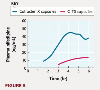
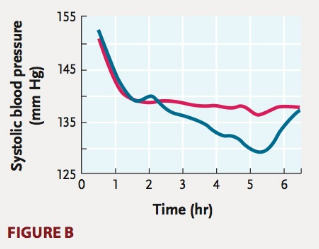
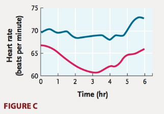
Nifedipine is an antihypertension medication that is also used to reduce the workload on the heart. The study below compares the effects of two different types of capsules used to administer the drug to patients (GITS—red line, and Cotracten X—blue line). Each graph shows changes with time following initial administra- tion of the capsules. Figure A compares the levels of nifedipine in the blood with each capsule type. The ability of the different cap- sules to reduce workload on the heart by altering blood pressure or heart rate is shown in Figures B and C, respectively.
Which type of capsule would you expect to have fewer side effects?
Source: M. J. Brown and C. B. Toal. 2008. Formulation of long-acting nifedipine tablets influences the heart rate and sympathetic nervous system response in hypertensive patients. British Journal of Clinical Pharmacology 65:646–652.

Trending nowThis is a popular solution!

Chapter 44 Solutions
Biology: The Dynamic Science (MindTap Course List)


- Nifedipine is an antihypertension medication that is also used to reduce the workload on the heart. The study below compares the effects of two different types of capsules used to administer the drug to patients (GITSred line, and Cotracten Xblue line). Each graph shows changes with time following initial administra- tion of the capsules. Figure A compares the levels of nifedipine in the blood with each capsule type. The ability of the different cap- sules to reduce workload on the heart by altering blood pressure or heart rate is shown in Figures B and C, respectively. Are elevated plasma levels of nifedipine(above 10 ng/mL) required for the drug to perform its actions? Source: M. J. Brown and C. B. Toal. 2008. Formulation of long-acting nifedipine tablets influences the heart rate and sympathetic nervous system response in hypertensive patients. British Journal of Clinical Pharmacology 65:646652.arrow_forwardWhat can you say about the amplitude of the various waves in different cardiac cycles? The P wave and the QRS complex represent depolarization of the atrial and ventricular muscle respectively. Why does the QRS complex have the largest amplitude? In Steps 7 and 8, heart rate was calculated based upon the peak-to-peak interval of the R waves. Was there variability between the beats? Would you expect the interval between beats to be identical? Why or why not? The range for a normal resting heart rate is 60 to 90 bpm. A trained athlete could have a resting heart rate of 45 to 60 bpm. Why might a very fit person have a slower heart rate than someone of average fitness? Are the amplitudes and durations of the various waves in different individuals similar or very different? What variations in heart rate did you observe between individuals? Explain why ventricular contraction (systole) and the ‘lub’ sound occur immediately after the QRS complex. Explain why ventricular relaxation…arrow_forwardThe below graph shows changes in several cardiovascular parameters as the result of increased intensity of exercise in a population of untrained individuals. What commonly measured cardiovascular parameters might the curves labeled A, B, and C represent? What are the corresponding units for the y-axis of each measure? For your reference, the two labeled curves represent the Pulmonary Capillary Wedge Pressure (PCWP) and the Central Venous Pressure (CVP), both of which are measured in mmHg.arrow_forward
- Give a detailed mechanism of action of a name drug or agonist that results in positive inotropic changes in the cardiac cell.arrow_forwardHow does COPD correlate with left ventricular pressure and primary heart failure? How many patients are suffering from COPD in the United States? Do COPD sufferers die of respiratory causes or other causes? (Be sure to cite the data.)arrow_forwardMATCH the NUMBERS from m the heart diagram with the description: some numbers can be used more than once.arrow_forward
- Which of the choices below best describes the systemic circulation of blood?a. The flow of blood into the right atrium and eventually out of the left ventricle.b. The movement of blood from the pulmonary trunk, through capillaries into the pulmonary veins.c. The movement of blood into the coronary arteries, through capillaries into the coronary sinus.d. The movement of blood from the aorta, through arteries and capillaries and then eventually to the vena cavae.arrow_forwardAll of the following are correct about the isovolumetric contraction, except ____________. A. This phase of the cardiac cycle begins with the appearance of the QRS complex of the ECG, which represents atrial repolarization and ventricular depolarization B. It represents the time period between the closure of the AV valves and the opening of the aortic and pulmonic valves, ventricular pressure rises rapidly without a change in ventricular volume C. The rate of pressure increase in the ventricles is determined by the rate of contraction of the muscle fibers, which is determine by mechanisms governing excitation-contraction coupling D. Ventricular chamber geometry changes considerably as the heart becomes more spheroid in shape; circumference increases and atrial base-to-apex length increasesarrow_forwardWhich of the following sequences shows the correct pathway of blood from body to lungs? body-superior and inferior vena cava-right atrium-tricuspid valve-right ventricle-pulmonary-semilunar valve-pulmonary trunk-pulmonary arteries-lungs body-superior and inferior vena cava-left atrium-tricuspid valve-left ventricle-aortic semilunar valve-pulmonary trunk-pulmonary arteries-lungs body-superior and inferior vena cava-right atrium-bicuspid valve-right ventricle-pulmonary-semilunar valve-pulmonary trunk-pulmonary veins-lungs body-superior and inferior vena cava-left atrium-tricuspid valve-left ventricle-pulmonary semilunar valve- pulmonary trunk-pulmonary arteries-lungsarrow_forward
- Give biological reason for the following: the wall of the ventricle is thicker than that of auricle.arrow_forwardAnatomic relationships to the sector image are orientated so that ______-sided structures such as the RA and RV are seen at the _____ of the image while ______-sided structures such as the LA and LV are seen towards the __________ of the image; ____________ structures such as the apices of both ventricles appear towards the __________ of the image while ____________ structures such as the atria appear towards the _________ of the image.arrow_forwardExplain the principle behind the Frank–Starling law of the heart. How does this mechanism normally prevent pulmonary or systemic congestion?arrow_forward
 Biology: The Dynamic Science (MindTap Course List)BiologyISBN:9781305389892Author:Peter J. Russell, Paul E. Hertz, Beverly McMillanPublisher:Cengage Learning
 Human Physiology: From Cells to Systems (MindTap ...BiologyISBN:9781285866932Author:Lauralee SherwoodPublisher:Cengage Learning
 Human Biology (MindTap Course List)BiologyISBN:9781305112100Author:Cecie Starr, Beverly McMillanPublisher:Cengage LearningEssentials of Pharmacology for Health ProfessionsNursingISBN:9781305441620Author:WOODROWPublisher:Cengage