
Videos
Problem Set
QUANTITATIVE Limits of Resolution Then and Now. Based on what you learned in this chapter about the limit of resolution of a light microscope, answer each of the following questions. Assume that the unaided human eye has a limit of resolution of about 0.25 mm and that a modern light microscope has a useful magnification of about 1000×.
(a) Define limit of resolution in your own words. What was the limit of resolution of Hooke’s microscope? What about van Leeuwenhoek’s microscope?
(b) What are the approximate dimensions of the smallest structure that Hooke would have been able to observe with his microscope? Would he have been able to see any of the structures shown in Figure 1-3a? If so, which ones? And if not, why not?
(c) What are the approximate dimensions of the smallest structure that van Leeuwenhoek would have been able to observe with his microscope? Would he have been able to see any of the structures shown in Figure 1-3a? If so, which ones? And if not, why not?
(d) What are the approximate dimensions of the smallest structure that a contemporary cell biologist should be able to observe with a modern light microscope?
(e) Consider the eight structures shown in Figure 1-3a and 1-3b. Which of these structures would both Hooke and van Leeuwenhoek have been able to see with their respective microscopes? Which, if any, would van Leeuwenhoek have been able to see that Hooke could not? Explain your reasoning. Which, if any, that neither Hooke nor van Leeuwenhoek could see would a contemporary cell biologist be able to see using a modern light microscope?
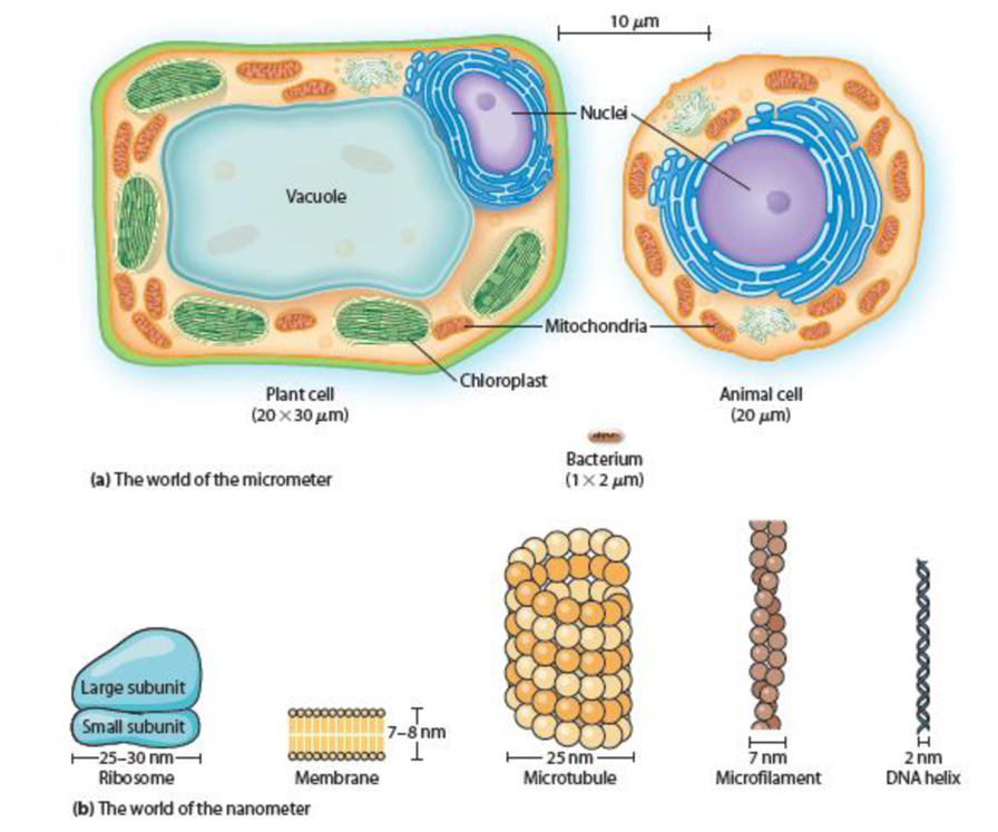
Figure 1-3 The Worlds of the Micrometer and Nanometer. Illustrations show (a) typical cells and (b) common cellular structures.
Want to see the full answer?
Check out a sample textbook solution
Chapter 1 Solutions
Becker's World of the Cell (9th Edition)


- Question:- Starting with data collection, describe the steps necessary to determine a 3-Dimensional structure using electron microscopy methods.arrow_forwardSDS-PAGE, gel electrophoresis: Is there a dye front on this gel? I am trying to measure relative mobility, but I am not sure of the final band on the ladder lane is an actual band or is the dye front. If it is an actual band and there is no dye front, where do I measure to in order to calculate relative mobilitiy (Rf)?arrow_forwardTrue or false? light microscopy has a limit of resolution of 200nm. phase contrast microscopy can be used to observ thick (more than 2 cells)speciments. mammalian cells are grown in a humidified incubator at 30C and 5% CO2.arrow_forward
- Question: Recommended tool for a plant protein (Scansite, NetPhos, GPS) Why?arrow_forwardPractice problems: Do on folder paper. 1. You are observing a cell; total magnification 100X. It looks like about 7 cells will fit across the field of view's diameter. How large is this cell? Explain. 2. For the 2nd specimen, you are using a total magnification of 400X. It's harder to estimate this time, so you approximated that 12 cells would fit across the radius. Explain how you would find out the size of one cell.arrow_forwardInstruction: FILL IN THE BLANKS. 1. In viewing objects under the microscope, it is observed that the microscope ______________ the direction of movement and _______________ the position of the image observed. 2. In shifting to higher magnification, simply rotate the _________________ to the desired objective. If the image is not clear manipulate the __________________ knob. 3. The total magnification of the specimen as seen in the microscope is 400x. This means that the ocular magnification is _______ x and the ______________ is 40x.arrow_forward
- True or false? each objective of a phase contrast microscope contains a phase plate. most cultured mammalian cells grow best at 37C. the oculars on the compound light microscope are typically 10X.arrow_forward1.Why is wavelenth the main limiting factor on limit of resoltuin in light microscopy? 2.Assuming that all other variables remain constant, explain why light of shorter wavelengths will produce a clearer image that light of longer wavelenths. 3. Why aren't the magnification of both ocular lenses of a binocular microscope used to calculate total magnification?arrow_forwardGolden Line:Rationale: (Tell me why the authors put this diagram here. What do you think is its main purpose for being in our textbook?)Personal Connection:Question:arrow_forward
- You may want to use this resource for this problem. If you do, submit the output along with your solution.You have been given a confocal microscope equipped with the following lasers, excitation filters, andemission filters:Laser Emission filter355 nm 410-470 nm405 nm 470-500 nm488 nm 500-550 nm532 nm 570-610 nm561 nm 610-650 nm640 nm 660-700 nm808 nm 720-780 nmYour task is to design an experiment to visualize the following:1. Nuclei2. A fluorescent protein in the cytosol3. A cell membrane marker antibody conjugated with a fluorophore4. Actin filaments5. LysosomesYou may choose from the following fluorophores for each of the five channels:Nuclei Fluorescent protein Membrane marker Actin marker Lysosome trackerDAPI GFP FITC AF488 Phalloidin LysoTracker RedHoechst 33342 YFP WGA-TRITC AF568 Phalloidin LysoTracker DeepRedSYTO Deep Red RFP Cy7 AF594 Phalloidin LysoTracker Blue Part 3.1Choose appropriate fluorophores for each of the subcellular structures to be imaged, taking into…arrow_forwardQuestion:- We are using in class 4.5 mm and 40X. a) How big is a specimen that takes up 1/2 the field view at 90X?arrow_forwardInstruction: FILL IN THE BLANKS 1. Manipulate the ________________ knob to bring the objective about 1 to 2 mm away from the slide. Never look into the ____________ when bringing the objective close to the slide to avoid damaging or crushing the slide. A clear field of vision should be ___________ not gray. 2. In viewing objects under the microscope, it is observed that the microscope ______________ the direction of movement and _______________ the position of the image observed. 3. In shifting to higher magnification, simply rotate the _________________ to the desired objective. If the image is not clear manipulate the __________________ knob. 4. The total magnification of the specimen as seen in the microscope is 400x. This means that the ocular magnification is _______ x and the ______________ is 40x.arrow_forward
 Human Anatomy & Physiology (11th Edition)BiologyISBN:9780134580999Author:Elaine N. Marieb, Katja N. HoehnPublisher:PEARSON
 Biology 2eBiologyISBN:9781947172517Author:Matthew Douglas, Jung Choi, Mary Ann ClarkPublisher:OpenStax
 Anatomy & PhysiologyBiologyISBN:9781259398629Author:McKinley, Michael P., O'loughlin, Valerie Dean, Bidle, Theresa StouterPublisher:Mcgraw Hill Education,
 Molecular Biology of the Cell (Sixth Edition)BiologyISBN:9780815344322Author:Bruce Alberts, Alexander D. Johnson, Julian Lewis, David Morgan, Martin Raff, Keith Roberts, Peter WalterPublisher:W. W. Norton & Company
 Laboratory Manual For Human Anatomy & PhysiologyBiologyISBN:9781260159363Author:Martin, Terry R., Prentice-craver, CynthiaPublisher:McGraw-Hill Publishing Co.
 Inquiry Into Life (16th Edition)BiologyISBN:9781260231700Author:Sylvia S. Mader, Michael WindelspechtPublisher:McGraw Hill Education