To analyze:
The major bones of the skeleton.
Introduction:
The internal framework of the body is provided by the human skeleton. The skeleton is composed of appendicular and axial skeleton. The axial skeleton includes rib cage, vertebral column, and skull. The shoulder girdle, pelvic girdle and limb bones together forms the appendicular skeleton.
Explanation of Solution
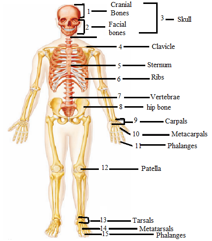
The skull is composed of facial and cranial bones. The bone present in the top part of the skull is the cranial bone that protects the brain. The cranial bone consist of parietal, frontal, sphenoid, ethmoid and occipital. The facial bones include maxilla, mandible, nasal bones, zygoma and frontal bone. Facial bones forms the structure of the face. The bone present between the sternum and the shoulder blade is the collar bone also called clavicle. This is the only horizontally lying bone in the body. The central part of the chest is occupied by a bone called chest bone. This bones connects the cartilage with the ribs. There are 24 ribs present in the body that protects the thoracic cavity.
The thirty three segmented vertebrae together forms the spine or the vertebral column. The vertebral column provides stiffness to the body. The large irregular bone present in the center of the body is the hip bone. This bone allows body to sit, and support the weight of the body. The hand consist of three types of bones that are phalanges, metacarpal and carpal bones. The phalanges are present in the fingers of the hands and toes.
The middle part of the hand consist of metacarpal bones. The wrist is composed of the bones called carpal bones. The kneecap is called the patella, a flat triangular bone that articulates with the femur. The patella provides protection and covers the articular surface of the knee joint. The bones in the foot are tarsals, metatarsals and phalanges. The ankle of the foot consist of bone called tarsals. The metatarsals are the bones that connects the tarsals and phalanges.
Want to see more full solutions like this?
Chapter 13 Solutions
Laboratory Manual for Holes Human Anatomy & Physiology Fetal Pig Version

 Human Anatomy & Physiology (11th Edition)BiologyISBN:9780134580999Author:Elaine N. Marieb, Katja N. HoehnPublisher:PEARSON
 Biology 2eBiologyISBN:9781947172517Author:Matthew Douglas, Jung Choi, Mary Ann ClarkPublisher:OpenStax
 Anatomy & PhysiologyBiologyISBN:9781259398629Author:McKinley, Michael P., O'loughlin, Valerie Dean, Bidle, Theresa StouterPublisher:Mcgraw Hill Education,
 Molecular Biology of the Cell (Sixth Edition)BiologyISBN:9780815344322Author:Bruce Alberts, Alexander D. Johnson, Julian Lewis, David Morgan, Martin Raff, Keith Roberts, Peter WalterPublisher:W. W. Norton & Company
 Laboratory Manual For Human Anatomy & PhysiologyBiologyISBN:9781260159363Author:Martin, Terry R., Prentice-craver, CynthiaPublisher:McGraw-Hill Publishing Co.
 Inquiry Into Life (16th Edition)BiologyISBN:9781260231700Author:Sylvia S. Mader, Michael WindelspechtPublisher:McGraw Hill Education