
Concept explainers
To review:
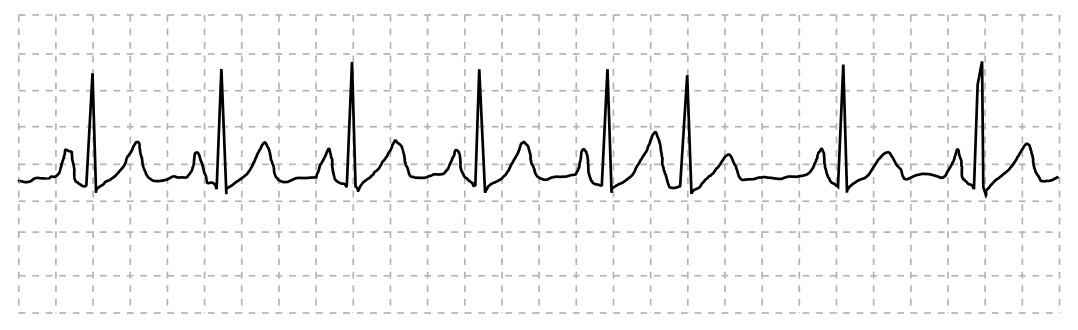
The given blank space in the statement using the following ECG graph.
QRS duration: __________ QT duration: ____________
Ventricular rate and rhythm: _____________
Atrial rate rhythm: _____________________
PR interval: __________________________
Interpretation: ________________________
Introduction:
The electrocardiogram or ECG is a tool that is used to record a patient’s heart rate in real time. The heartbeat produces electrical signals that are recorded in this tool. It is recorded in a waveform, which has P, Q, R, S, T, and U components. The ECG is interpreted by doctors to know the condition of patients.
Explanation of Solution
The heart rate of the patient is regular with normal electrical activity of the heart. There was one PAC (premature atrial contractions), also known as atrial premature beats, at sixth position, which occurred naturally. The ECG tracing is interpreted as normal sinus rhythm.
QRS duration: The duration of the QRS complex is less than 0.12 seconds.
QT duration: It is more than 0.12 seconds.
Ventricular rate and rhythm: The ventricular rate is regular and between 60 and 100bpm. Ventricular rhythm is less than 0.12 seconds.
Atrial rate rhythm: It is regular with the heart rate up to 100 bpm and occurrence of one PAC at the 6th complex.
PR interval: Duration is less than 0.20 seconds.
Interpretation: The patient’s heart rate is between 60 and 100 bpm, so the graph represents ECG tracing of Normal Sinus Rhythm (NSR) with one PAC at the sixth complex.
QRS duration: 0.6 seconds QT duration: 0.40 seconds
Ventricular rate and rhythm: 86 bpm
Atrial rate rhythm: 86 bpm
PR interval: 0.16 seconds
Interpretation: The graph shows normal sinus rhythm with 86 bpm having one atrial premature beat at the sixth complex
Want to see more full solutions like this?
Chapter 14 Solutions
MindTap for Des Jardins' Cardiopulmonary Anatomy & Physiology, 2 terms Printed Access Card


- Why is percussion omitted in heart assessment?arrow_forward(a) Referring to the figure, find the time systolic pressure lags behind the middle of the QRS complex. (b) Discuss the reasons for the time lag.arrow_forwardIn electrocardiography (=EKG, =ECG), what causes a P wave?______ A. atrial repolarization B. ventricular repolarization C. atrial depolarization D. ventricular depolarizationarrow_forward
- The small squares on the standard ECG paper representA. 0.02 secondB. 0.04 secondC. 0.06 secondD. 0.08 secondarrow_forwardThe standard EKG consists of 10 sensors that record 12 leads of the heart’s electrical activity from different angles, allowing for a thorough three-dimensional interpretation of its activity. This is transmitted by the electrodes to the equipment to be interpreted and is used to diagnose cardiac medical conditions. In case of an abnormal EKG, the second step would be to use a Holter monitor. How would you explain how to perform an EKG (steps)? Where will you place the electrodes when performing and EKG? Why? What are the different lead types, connections, and placements? When you conclude an EKG, what are the different components that you need to observe and confirm before you disconnect the patient? Can you explain the difference between normal, abnormal, and artifacts? What is a Holter monitor? Under what circumstances would one be ordered for a patient? How do you use a Holter monitor? Educate a patient: What you will do before, during, and after an electrocardiogram or…arrow_forward
 Cardiopulmonary Anatomy & PhysiologyBiologyISBN:9781337794909Author:Des Jardins, Terry.Publisher:Cengage Learning,
 Basic Clinical Lab Competencies for Respiratory C...NursingISBN:9781285244662Author:WhitePublisher:Cengage
- Understanding Health Insurance: A Guide to Billin...Health & NutritionISBN:9781337679480Author:GREENPublisher:Cengage
 Human Physiology: From Cells to Systems (MindTap ...BiologyISBN:9781285866932Author:Lauralee SherwoodPublisher:Cengage Learning