Laboratory Manual for Human Anatomy & Physiology Main Version
4th Edition
ISBN: 9781260159110
Author: Terry Martin
Publisher: Mcgraw-hill Higher Education (us)
expand_more
expand_more
format_list_bulleted
Videos
Textbook Question
Chapter 15, Problem F15.11A
FIGURE 15.11 Label the bones and features of the thoracic cage, using the terms provided 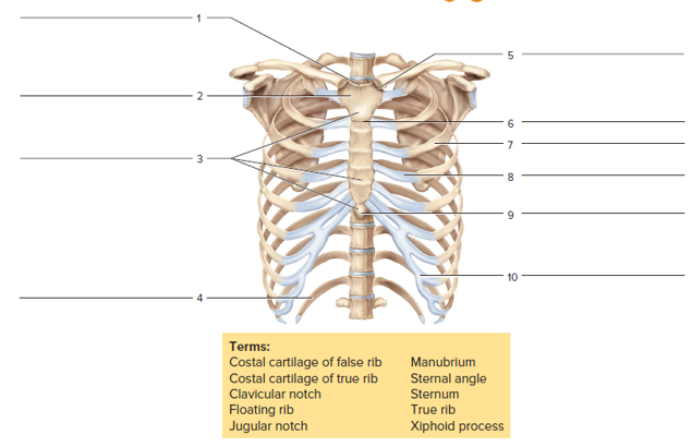
Expert Solution & Answer
Want to see the full answer?
Check out a sample textbook solution
Students have asked these similar questions
Identify the labeled parts of the vertebrae
identify the following bones and features of the axial skeleton
name the features(red circles)
Write the name and numbers of the bones of thoracic cage. Which bones form the superior thoracic aperture
Chapter 15 Solutions
Laboratory Manual for Human Anatomy & Physiology Main Version
Ch. 15 - The most superior bone of the vertebral column is...Ch. 15 - The vertebral column possesses four curvatures....Ch. 15 - Humans have ___________ pairs of true ribs. two...Ch. 15 - The _________ ribs do not have costal cartilage...Ch. 15 - Humans possess ____________ cervical vertebrae....Ch. 15 - The superior end of the sacrum articulates with...Ch. 15 - The anterior (sternal) end of a rib articulates...Ch. 15 - All cervical, thoracic, and lumbar vertebrae...Ch. 15 - A feature of the second cervical vertebra is the...Ch. 15 - Note the four curvatures of the vertebral column....
Ch. 15 - The vertebral column encloses and protects the...Ch. 15 - The vertebral column extends from the skull to the...Ch. 15 - The seventh cervical vertebra is called the...Ch. 15 - The _____________________ of the vertebrae support...Ch. 15 - The __________ separate adjacent vertebrae, and...Ch. 15 - The intervertebral foramina provide passageways...Ch. 15 - Transverse foramina of _____________ vertebrae...Ch. 15 - The first vertebra also is called the...Ch. 15 - When the head is moved from side to side, the...Ch. 15 - The __________ vertebrae have the largest and...Ch. 15 - The typical number ofvertebrae that fuse in the...Ch. 15 - FIGURE 15.8 Label the bones and features of a...Ch. 15 - FIGURE 15.9 Identity the bones and features...Ch. 15 - An abnormal lateral curvature of the spine is...Ch. 15 - The manubrium, body, and xiphoid process form a...Ch. 15 - The last two pairs of fibs that have no...Ch. 15 - There are _____________ pairs of true ribs.Ch. 15 - Costal are composed of __________ tissue.Ch. 15 - The manubriunarticulates with _________ on its...Ch. 15 - List three general functions ofthe thoracic cage.Ch. 15 - The sternal angle indicates the location of the...Ch. 15 - FIGURE 15.11 Label the bones and features of the...
Additional Science Textbook Solutions
Find more solutions based on key concepts
Figure 1.18 In the example below, the scientific method is used to solve an everyday problem. Which part in the...
Concepts of Biology
Which of the following would be used to identify an unknown bacterial culture that came from a patient in the i...
Microbiology Fundamentals: A Clinical Approach
Problem Set
True or False? Indicate whether each of the following statements about membrane transport is true (...
Becker's World of the Cell (9th Edition)
What were the major microbiological interests of Martinus Beijerinck and Sergei Winogradsky? It can be said tha...
Brock Biology of Microorganisms (15th Edition)
a. What three lineages of lobe-fins survive today? b. Go back to the phylogenetic tree in Interactive Question ...
Study Guide for Campbell Biology
Knowledge Booster


Learn more about
Need a deep-dive on the concept behind this application? Look no further. Learn more about this topic, biology and related others by exploring similar questions and additional content below.Similar questions
- Name the bones that form the upper limb, from proximal to distal. 8.2 Upper Limb (Extremity)arrow_forwardSpecimen: Chicken Bones Lumbar and sacral vertebrae: There are several bones in synsacrum (made up of thoracic, lumbar, and sacral vertebrae). Describe the synsacrum in your specimen.arrow_forwardIdentify four (4) major regions of the vertebral column. Describe how the vertebrae located in the neck differ from the vertebrae in the lower backarrow_forward
- identify the following bones of the axial skeleton and write the answers belowarrow_forwardUse the labels from figure 13.1 and color in the bones of the upper extremity in this anterior view. The inset is a posterior view of the elbow jointarrow_forwardFigure 7b.7. The left orbit, anterior view. (a) Structures of the orbit. (b) Photograph of the orbit to be labeled. 5. 2. SUPIOOYBHA maYgin * Z49omatlC 1acrimal BOne OPTIC Canal 7. SUPEKIOK OPDilal riEsuet पाम्बनाप . 6.arrow_forward
- The image shows the last 2 thoracic vertebrae of DOG (T12,T13) in lateral view. Find and label the following: Arch Body Spinous process Caudal articular process Cranial articular process Mammillary process Accessory process Fovea of transverse process Cranial costal fovea Lamina Pediclearrow_forwardIdentify the bones and bone markings in Figurearrow_forwardComplete the blank spaces ( name of joints)arrow_forward
arrow_back_ios
SEE MORE QUESTIONS
arrow_forward_ios
Recommended textbooks for you

 Medical Terminology for Health Professions, Spira...Health & NutritionISBN:9781305634350Author:Ann Ehrlich, Carol L. Schroeder, Laura Ehrlich, Katrina A. SchroederPublisher:Cengage Learning
Medical Terminology for Health Professions, Spira...
Health & Nutrition
ISBN:9781305634350
Author:Ann Ehrlich, Carol L. Schroeder, Laura Ehrlich, Katrina A. Schroeder
Publisher:Cengage Learning
The Skeletal System; Author: Professor Dave Explains;https://www.youtube.com/watch?v=f-FF7Qigd3U;License: Standard YouTube License, CC-BY