
LAB MANUAL FOR HUMAN A&P MAIN VERSION
4th Edition
ISBN: 9781266871016
Author: Martin
Publisher: MCG
expand_more
expand_more
format_list_bulleted
Videos
Textbook Question
Chapter 15, Problem F15.9A
FIGURE 15.9 Identity the bones and features indicated in this radiograph of the neck (lateral view), using the terms provided.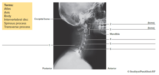
Expert Solution & Answer
Want to see the full answer?
Check out a sample textbook solution
Students have asked these similar questions
When the head is moved from side to side, the first vertebra pivots around the _______________________ of thesecond vertebra.
From the given Left nasal bone (dog), ventralateral view, locate the:
Median border
Caudal border
Lateral border
Specimen: Chicken Bones
Lumbar and sacral vertebrae: There are several bones in synsacrum (made up of thoracic, lumbar, and sacral vertebrae). Describe the synsacrum in your specimen.
Chapter 15 Solutions
LAB MANUAL FOR HUMAN A&P MAIN VERSION
Ch. 15 - The most superior bone of the vertebral column is...Ch. 15 - The vertebral column possesses four curvatures....Ch. 15 - Humans have ___________ pairs of true ribs. two...Ch. 15 - The _________ ribs do not have costal cartilage...Ch. 15 - Humans possess ____________ cervical vertebrae....Ch. 15 - The superior end of the sacrum articulates with...Ch. 15 - The anterior (sternal) end of a rib articulates...Ch. 15 - All cervical, thoracic, and lumbar vertebrae...Ch. 15 - A feature of the second cervical vertebra is the...Ch. 15 - Note the four curvatures of the vertebral column....
Ch. 15 - The vertebral column encloses and protects the...Ch. 15 - The vertebral column extends from the skull to the...Ch. 15 - The seventh cervical vertebra is called the...Ch. 15 - The _____________________ of the vertebrae support...Ch. 15 - The __________ separate adjacent vertebrae, and...Ch. 15 - The intervertebral foramina provide passageways...Ch. 15 - Transverse foramina of _____________ vertebrae...Ch. 15 - The first vertebra also is called the...Ch. 15 - When the head is moved from side to side, the...Ch. 15 - The __________ vertebrae have the largest and...Ch. 15 - The typical number ofvertebrae that fuse in the...Ch. 15 - FIGURE 15.8 Label the bones and features of a...Ch. 15 - FIGURE 15.9 Identity the bones and features...Ch. 15 - An abnormal lateral curvature of the spine is...Ch. 15 - The manubrium, body, and xiphoid process form a...Ch. 15 - The last two pairs of fibs that have no...Ch. 15 - There are _____________ pairs of true ribs.Ch. 15 - Costal are composed of __________ tissue.Ch. 15 - The manubriunarticulates with _________ on its...Ch. 15 - List three general functions ofthe thoracic cage.Ch. 15 - The sternal angle indicates the location of the...Ch. 15 - FIGURE 15.11 Label the bones and features of the...
Additional Science Textbook Solutions
Find more solutions based on key concepts
Your bore cells, muscle cells, and skin cells look different because a. different kinds of genes are present in...
Campbell Essential Biology (7th Edition)
Describe the evolution of mammals, tracing their synapsid lineage from early amniote ancestors to true mammals....
LooseLeaf for Integrated Principles of Zoology
A student moving out of a dormitory crouches in correct fashion to lift a heavy box of books. What prime movers...
HUMAN ANATOMY
The appearance of glucose in the urine a. occurs normally. b. indicates the presence of kidney disease. c. occu...
Human Physiology
a. What three lineages of lobe-fins survive today? b. Go back to the phylogenetic tree in Interactive Question ...
Study Guide for Campbell Biology
Knowledge Booster


Learn more about
Need a deep-dive on the concept behind this application? Look no further. Learn more about this topic, biology and related others by exploring similar questions and additional content below.Similar questions
- Identify the structures in Figurearrow_forwardThe image shows the last 2 thoracic vertebrae of DOG (T12,T13) in lateral view. Find and label the following: Arch Body Spinous process Caudal articular process Cranial articular process Mammillary process Accessory process Fovea of transverse process Cranial costal fovea Lamina Pediclearrow_forwardLabel the bones of the hand in Figure. For each phalanx, include the Roman numeral.asaparrow_forward
- Figure 7b.7. The left orbit, anterior view. (a) Structures of the orbit. (b) Photograph of the orbit to be labeled. 5. 2. SUPIOOYBHA maYgin * Z49omatlC 1acrimal BOne OPTIC Canal 7. SUPEKIOK OPDilal riEsuet पाम्बनाप . 6.arrow_forwardThe temporal bones are joined to the parietal bones along the _______________________ sutures.arrow_forwardLabel the bones of the hand in Figure. For each phalanx, include the Roman numeral. Asap quicklyarrow_forward
- Please label the frog skeleton accordinglyarrow_forwardLabel the bony structures of the shoulder and upper limb. Infraspinous fossa Anatomical neck Supraspinous fossa Inferior angle Olecranon Medial border Lateral border Surgical neckarrow_forwardSpecimen: Chicken Bones In thoracic vertebrae, observe if the vertebrae are fused or not. Observe its characteristics and relate to its function in a living chicken.arrow_forward
arrow_back_ios
SEE MORE QUESTIONS
arrow_forward_ios
Recommended textbooks for you
 Medical Terminology for Health Professions, Spira...Health & NutritionISBN:9781305634350Author:Ann Ehrlich, Carol L. Schroeder, Laura Ehrlich, Katrina A. SchroederPublisher:Cengage Learning
Medical Terminology for Health Professions, Spira...
Health & Nutrition
ISBN:9781305634350
Author:Ann Ehrlich, Carol L. Schroeder, Laura Ehrlich, Katrina A. Schroeder
Publisher:Cengage Learning
The Skeletal System; Author: Professor Dave Explains;https://www.youtube.com/watch?v=f-FF7Qigd3U;License: Standard YouTube License, CC-BY