
Videos
Connecting the Concepts
1. There are several key concepts introduced in this chapter: Structure correlates with function: an animal’s body has a hierarchy of organization with emergent properties at each level: and complex bodies have structural adaptations that increase surface area for exchange. Label the tissue layers shown in this section of the small intestine, and describe how this diagram illustrates these three concepts.
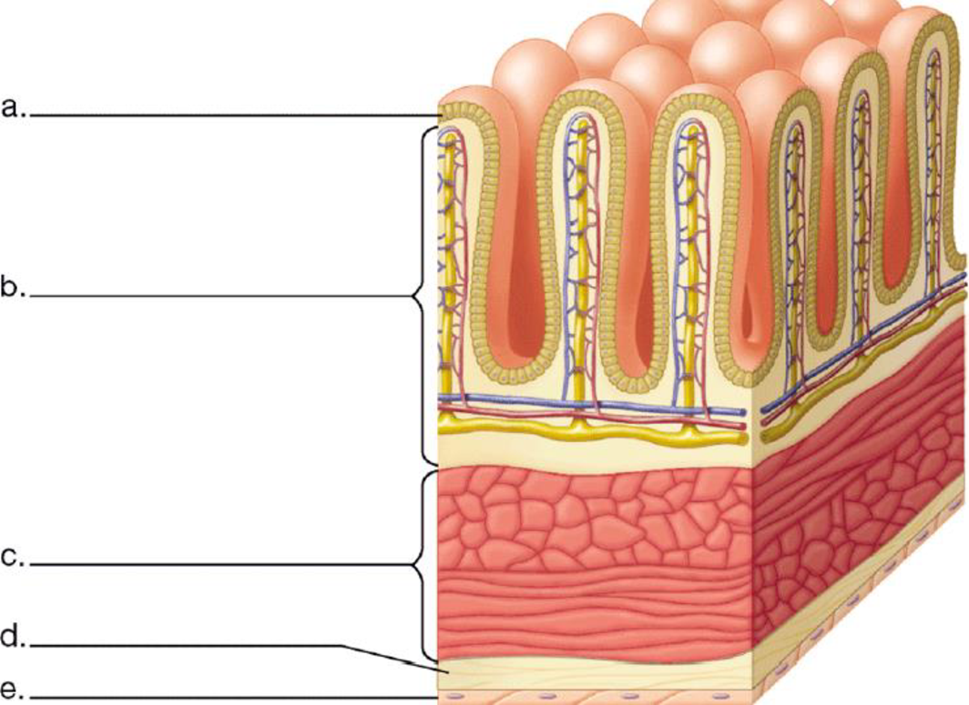
To label: The tissue layers as shown in the section of small intestine. Also to describe the concept that ‘the structure correlates with functions’; ‘an animal’s body has a hierarchy of organization with emergent properties at each level’; and ‘complex bodies have structural adaptations that increase surface area for exchange’ in the diagram.
Introduction: The structure of each specialized cell type fits to their functions. A tissue derives its function by the cells that make it up. For example,the columnar epethelial cells.
Connective tissues have fibres and cells that provide support and connection to the tissue. Therefore, the connection of cell to tissue to the organ is visible in the given diagram.
There are many projections that line the surface of the small intestine for the absorption of the nutrients. The villi increases the surface area of the small intetine for absorption of nutrients.
Answer to Problem 1CC
Pictorial representation: A labeled diagram showing different tissue layers in a section of small intestine is presented in the Fig.1.
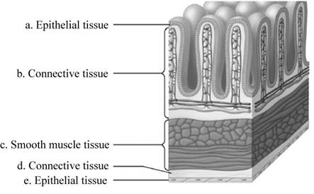
Fig.1: Different section of small intestine.
Explanation of Solution
(a)
Correct answer: Epithelial tissue.
Explanation: In the small intestine, epithelial tissue covers the outside of the villi. Hence the correct answer is epithelial tissue.
(b)
Correct answer: Connective tissue.
Explanation: Connective tissue attaches and provides support to the organs. Hence the correct answer is connective tissue
(c)
Correct answer: Smooth muscle tissue.
Explanation: Small intestine requires strength to push the digested product down its length, and this is provided by smooth muscle tissue. Hence the correct answer is smooth muscle tissue.
(d)
Correct answer: Connective tissue.
Explanation: Connective tissue attaches and provides support to the organs. Hence the correct answer is connective tissue.
(e)
Correct answer: Epithelial tissue.
Explanation: It attaches to the smooth muscle tissue and provide strength to the small intestine. Hence the correct answer is epithelial tissue.
Want to see more full solutions like this?
Chapter 20 Solutions
BSC 1005 PKG-W/MOD. MAST. ACCESS >CI<


- pig Trace the path of food through the pig’s digestive system. Explain the function of each organ as it contributes to the digestion of food.arrow_forwardCorrelate the structural changes in each segment of the digestive tract with the function. (Frog’s digestive system)arrow_forwardComposition of the Digestive System of Rabbitarrow_forward
- Answer the following questions about a Vegetarian diet: 1. Which major organ(s) is/are directly affected by the diet? 2. If applicable, what specific tissue or organelle would be affected by the diet. If no research is available, hypothesize which tissue or organelle may be affected and why. 3. Which nutrient (macro or micro) is the focus of this diet? 4. List the foods that would contain the nutrients that are the main focus of the diet. 5. List Pros and Cons of being on the diet. 6. List any possible misconceptions that may have been verified or nullified by scientific articles for this diet. 7. What biochemical pathway would this diet target? 8. List the sites that you use as references.arrow_forwardFetal Pig Oral Cavity Please refer to the pic to answer the question Describe the color, texture, and functions of the fetal pig's oral cavity. Compare the features of the pig's oral cavity to that of the frogs. What features of the pigs oral cavity are also found in that of humans? What differences are there? Describe the pigs teeth. Also what prevents food from entering the glottis or nasopharynx and what happens when the food does enter either of those two openings?arrow_forwardI. Give the derivatives of the following structures (one each only): 1. foregut - ____________________ 2. neural tube - _________________ 3. pharyngeal pouch I - ___________ 4. sclerotome - __________________ 5. laryngotracheal groove - ________ 6. telencephalon - _______________ 7. hepatic diverticulum - __________ 8. metencephalon - ______________ 9. cardiac neural crest - __________ 10. myotome - __________________ 11. lateral nasal - _______________ 12. hindgut - ___________________ 13. midgut - ___________________ 14. apaxial mesoderm - __________ 15. syndetome - ________________ 16. diencephalon - ______________ 17. dermatome - ________________ 18. optic vesicles - ______________ 19. meningotome - ______________ 20. pharyngeal pouch II - ________arrow_forward
- 1. compare the dissected pig human models viewed in the lab book structure pig human number of liver lobes location and shape of large intestine colon presence of appendix presence of cecumarrow_forwardDraw a plan diagram of the frog intestine viewed under the microscope (see attached images for help!!).... A plan diagram is a simple drawing showing only the boundaries between the lumen, the columnar epithelium, the (areolar) connective tissue, the circular smooth muscle and the longitudinal smooth muscle. A plan diagram serves to illustrate the location and relative thickness of the various tissue layers. 1. Draw a plan diagram and label the following: • Longitudinal smooth muscle • Columnar epithelial tissue • Circular smooth muscle • Villi • Areolar connective tissue • Lumen 2. Calculate actual size and drawing magnification of the width of the intestine. Include formulae used and calculations. 3. Add a scale bar with actual size next to your diagram. Give your drawing a descriptive title and record total magnification. Attached are an example plan diagram labelled, as well as a microscopic image of the frog intestine (3-4 cells should fit across only) PLEASE DO THE DRAWING AND…arrow_forwardWhich if the following is not a function of the digestive system 1. to ingest food 2. to digest food into small nutritions so that the molecules can pass through membrane 3. to absorb nutrient molecules 4. To eliminate indigestible remains 5. to deliver oxygen to Ty body tissuesarrow_forward
- Does the type of diet of the insect affect the structure of organs/parts of the digestive system? Cite at least four structures and explain such adaptation. Make sure that each division of the digestive system should be represented. Elaborate in your own words.arrow_forwardsea star oral 2.The _________________ ___________________ connect to each lobe of the digestive gland.arrow_forwardsea star oral 5.The ____________ __________________ is a circular tube that connects to each of the radial canals of each arm.arrow_forward
 Biology 2eBiologyISBN:9781947172517Author:Matthew Douglas, Jung Choi, Mary Ann ClarkPublisher:OpenStax
 Concepts of BiologyBiologyISBN:9781938168116Author:Samantha Fowler, Rebecca Roush, James WisePublisher:OpenStax College