Anatomy & Physiology (6th Edition)
6th Edition
ISBN: 9780134156415
Author: Elaine N. Marieb, Katja N. Hoehn
Publisher: PEARSON
expand_more
expand_more
format_list_bulleted
Concept explainers
Videos
Textbook Question
Chapter 20.6, Problem 18CYU
Class II MHC proteins display what kind of antigens? What class of T cell recognizes antigens bound to class II MHC? What types of cells display these proteins?
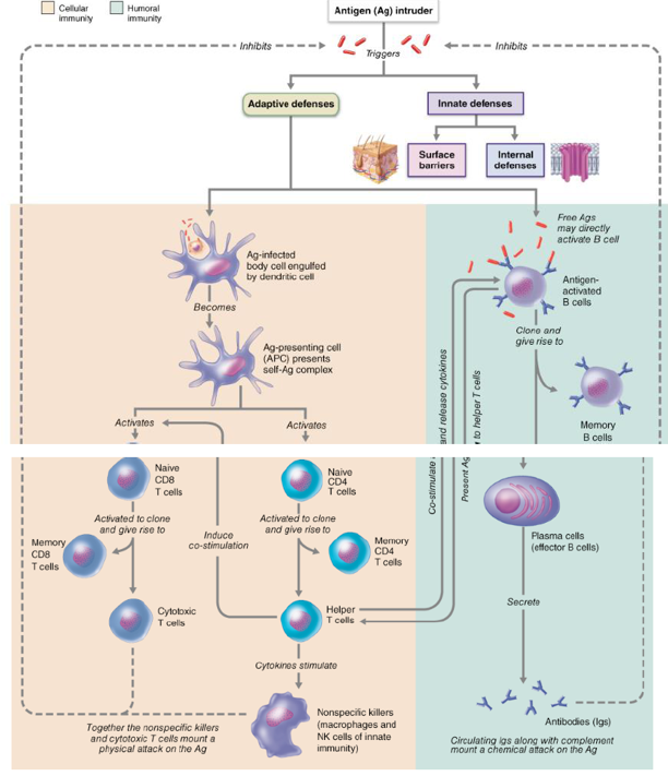
Figure 20.20 Simplified summary of the primary immune response.
Co-stimulation usually requires direct cell-cell interactions; cytokines enhance these and many other events. Although complement, NK cells, and phagocytes are innate defenses, they are enlisted in the fight by cytokines. (For simplicity, only B cell receptors are illustrated.)
Expert Solution & Answer
Want to see the full answer?
Check out a sample textbook solution
Students have asked these similar questions
Generally speaking, what kinds of cells express MHC I, MHC II, or both? What is presented on MHC I and II, and what kind of T cell recognizes each one? What response occurs when a T cell recognizes what is being presented on MHC I? What happens if it recognizes MHC II? Give an example of each type of recognition.
In regard to antigen presentation, MHC class I molecules usually present peptides derived from _____, whereas MHC class II molecules usually present peptides derived from _____.
a. intracellular cytosolic sources; vesicular system
b. phagolysosome; proteasomes
c. MIIC; self proteins
d. CLIP; HLA-DM
e. endocytic vesicles; endoplasmic reticulum.
Opsonization of pathogens by both antibodies and complement proteins (C3b) leads to uptake and destruction of the pathogen by phagocytic cells that express both Fc receptors and complement receptors. Which of the following in the figure below is the most efficient form of dual opsonization of the pathogen by antibody and C3b to maximize phagocytosis?
Chapter 20 Solutions
Anatomy & Physiology (6th Edition)
Ch. 20.1 - What distinguishes the innate defense system from...Ch. 20.1 - What is the first line of defense against disease?Ch. 20.2 - What is opsonization and how does it help...Ch. 20.2 - Under what circumstances might NK cells kill our...Ch. 20.2 - What are the cardinal signs of inflammation and...Ch. 20.3 - Name three key characteristics of adaptive...Ch. 20.3 - What is the difference between a complete antigen...Ch. 20.3 - What marks a cell as self as opposed to nonselfCh. 20.4 - What event (or observation) signals that a B or T...Ch. 20.4 - Which of the following T cells would survive...
Ch. 20.4 - Prob. 11CYUCh. 20.4 - In clonal selection, who does the selecting? What...Ch. 20.5 - Why is the secondary response to an antigen so...Ch. 20.5 - Prob. 14CYUCh. 20.5 - Which class of antibody is most abundant in blood?...Ch. 20.5 - List four ways in which antibodies can bring about...Ch. 20.5 - Prob. 17CYUCh. 20.6 - Class II MHC proteins display what kind of...Ch. 20.6 - Which type of T cell is the most important in both...Ch. 20.6 - Describe the killing mechanism of cytotoxic T...Ch. 20.7 - Prob. 21CYUCh. 20.7 - Prob. 22CYUCh. 20 - All of the following are considered innate body...Ch. 20 - The process by which neutrophils squeeze through...Ch. 20 - Antibodies released by plasma cells are involved...Ch. 20 - Which of the following antibodies can fix...Ch. 20 - Which antibody class is abundant in body...Ch. 20 - Small molecules that must combine with large...Ch. 20 - Lymphocytes that develop immunocompetence in the...Ch. 20 - Cells that can directly attack target cells...Ch. 20 - Prob. 9MCCh. 20 - The cell type most often invaded by HIV is a(n)...Ch. 20 - Complement fixation promotes all of the following...Ch. 20 - Using the letters from column B, match the cell...Ch. 20 - Besides acting as mechanical barriers, the skin...Ch. 20 - Explain why attempts at phagocytosis are not...Ch. 20 - What is complement? How does it cause bacterial...Ch. 20 - Interferons are referred to as antiviral proteins....Ch. 20 - Differentiate between humoral and cellular...Ch. 20 - Although the adaptive immune system has two arms,...Ch. 20 - Define immunocompetence and self-tolerance. How is...Ch. 20 - Differentiate between a primary and a secondary...Ch. 20 - Prob. 21SAQCh. 20 - What is the role of the variable regions of an...Ch. 20 - Name the five antibody classes and describe where...Ch. 20 - How do antibodies help defend the body?Ch. 20 - Do vaccines produce active or passive humoral...Ch. 20 - Prob. 26SAQCh. 20 - Describe the specific roles of helper, regulatory,...Ch. 20 - Prob. 28SAQCh. 20 - Prob. 29SAQCh. 20 - What events can result in autoimmune disease?Ch. 20 - Prob. 1CCSCh. 20 - Prob. 2CCSCh. 20 - Prob. 3CCSCh. 20 - Prob. 4CCSCh. 20 - Remember Mr. Ayers, the bus driver from Chapter...
Knowledge Booster


Learn more about
Need a deep-dive on the concept behind this application? Look no further. Learn more about this topic, biology and related others by exploring similar questions and additional content below.Similar questions
- Figure 42.11 Which of the following statements about T cells is false? Helper T cells release cytokines while cytotoxic T cells kill the infected cell. Helper T cells are CD4+, while cytotoxic T cells are CD8+. MHC II is a receptor found on most body cells, while MHC I is a receptor found on immune cells only. The T cell receptor is found on both CD4+ and CD8+ T cells.arrow_forwardHelper T cells are affected by HIV, how come is this receptor key to the immune system? which line of defense are we referring to? How is it connected to the immune system and which line of defense? Hence, based on your prompt, how are cytokines linked to the defense mechanism of HIV virus? Do you know or can you explain the cascade of events dealing with PAMPS, TLRs, interferon? What do they have to do with the second line of defense?arrow_forwardWhy is the concentration of this ligand for the NK-cell receptor CD94:NKG2A. on the target cell an effective measure of the presence or absence of classical MHC class I molecules?arrow_forward
- On which types of cells are the two classes of major histocompatibility complex (MHC) proteins located and what type of antigen do they display? MHC I proteins are found on the surface of antigen-presenting cells and display exogenous antigens, while MHC II proteins are found on the surface of most cells, and display endogenous antigens. MHC I proteins are found inside most cells, and display exogenous antigens, while MHC II proteins are found on the surface of antigen-presenting cells and display endogenous antigens. MHC I proteins are found inside all cells and display exogenous antigens, while MHC II proteins are found inside antigen-presenting cells and display endogenous antigens. MHC I proteins are found on the surface of most cells and display endogenous antigens, while MHC II proteins are only found on antigen-presenting cells and display exogenous antigens.arrow_forwardThe classical complement pathway is initiated by C1q binding to the surface of a pathogen. In some cases, C1q can directly bind the pathogen, for instance by recognizing proteins of bacterial cell walls, but in most cases C1q binds to IgM antibodies that are bound to the pathogen surface. How does this IgM-binding feature of C1q contribute to rapid, innate immune responses rather than to slow, adaptive responses? C1q induces B lymphocytes to begin secreting antibody within hours of pathogen exposure. Natural antibody that binds to many microbial pathogens is produced prior to pathogen exposure. C1q binds to C-reactive protein which then binds to IgM on the pathogen surface. C1q directly induces inflammation, recruiting phagocytes and antibodies from the blood into the infected tissue. C1q binds to dendritic cells in the infected tissue, inducing them to secrete inflammatory cytokines.arrow_forwardWhere are major histocompatibility complex-I (MHC-I) molecules located in the human body (cells)? Briefly describe how these surface markers present antigens to other cells. (Make sure to include where the antigen originates and what type of T-cell interacts with MHC-I antigen presentation.)arrow_forward
- Ingestion of complement-tagged pathogens by phagocytes is mediated by receptors for the bound complement proteins. Even when the complement cascade fails to proceed beyond generating the C3 convertase, complement activation is effective at inducing pathogen uptake and destruction. This process of immune protection is mediated by: Activation of complement inhibitory receptors on phagocytes that promote pathogen uptake Activation of soluble proteases in the serum that disrupt pathogen membranes Engagement of complement receptors on phagocytes by C3b and its cleavage products which promotes phagocytosis Engagement of complement receptors on B cells that promotes antibody production Stimulation of antimicrobial peptide secretion by phagocytesarrow_forwardWhich of the following statements is NOT correct? Group of answer choices -Natural killer cells are cytolytic lymphocytes that mediate ADCC -Neutrophils carry out phagocytosis of C3b-tagged particles -Macrophages carry out phagocytosis of C3b-tagged particles -Mast cells produce granules containing the cytotoxin perforin -B lymphocytes express a receptor for complement proteinsarrow_forwardB cells express a complement receptor that binds to C3b cleavage products, such as iC3b and C3dg. When a B cell with an antigen receptor that specifically recognizes that pathogen also has its complement receptor stimulated because the pathogen is opsonized with these C3 fragments, B cell activation is greatly enhanced. Due to this mechanism, B cells can be activated by much lower concentrations of antigen (in this case, the pathogen) than if the antigen is devoid of complement components. This mechanism functions to: Ensure that pathogens are readily detected by the adaptive immune system before they replicate to high levels in the host Prevent B cells from being activated in response to antigens that are not pathogens Allow B cells to phagocytose the pathogen and help destroy it Induce increased rounds of B cell replication to make more pathogen-specific B cells Allow the B cell to block pathogen replication by interfering with multiple pathogen surface functionsarrow_forward
- Three major cell types, dendritic cells, macrophages, and B cells, present peptides bound to MHC class II molecules for recognition by CD4 T cells. In general, these peptides are derived from proteins or pathogens taken up by the cell by endocytosis, phagocytosis, or macropinocytosis. Based on these pathways of antigen uptake, some of the enzymes that degrade proteins to generate peptides for MHC class II presentation are: Ubiquitin ligases that tag proteins for degradation by the proteasome ATP transporter proteins that deliver endocytic proteins into the cytosol for degradation Cysteine proteases like cathepsins that function at acidic pH The lysosomal thiol reductase found in the endosomes The lysosome-associated membrane trafficking protein, LAMP-2arrow_forwardSome viruses have mechanisms to down-regulate MHC class I protein expression on the surface of cells in which the virus is replicating. This immune evasion strategy might prevent effector CD8 cytotoxic T cells from recognizing and killing the virus-infected cells. Would this immune evasion strategy also prevent the initial activation of virus-specific CD8 T cells? Yes, because no viral peptide:MHC class I complexes would form to activate CD8 T cells. No, because dendritic cells would take up infected cells and cross-present viral peptides to activate CD8 T cells. No, because some presentation of MHC class I complexes with viral peptides would occur before the virus could down-regulate all the surface MHC class I protein. Yes, because this immune evasion strategy would also function in dendritic cells, even if the virus doesn’t replicate in dendritic cells. No, because the type I interferon response induced by the virus infection will up-regulate MHC class I expression and override the…arrow_forwardThe C1 complement protein is activated when it binds to the Fc region of an antigen-bound antibody. complement receptor of an antigen-bound lectin. Fc region of free antibody. complement receptor of free lectin. complement receptor on phagocytes.arrow_forward
arrow_back_ios
SEE MORE QUESTIONS
arrow_forward_ios
Recommended textbooks for you
 Biology 2eBiologyISBN:9781947172517Author:Matthew Douglas, Jung Choi, Mary Ann ClarkPublisher:OpenStax
 Human Physiology: From Cells to Systems (MindTap ...BiologyISBN:9781285866932Author:Lauralee SherwoodPublisher:Cengage Learning
Biology 2e
Biology
ISBN:9781947172517
Author:Matthew Douglas, Jung Choi, Mary Ann Clark
Publisher:OpenStax
Human Physiology: From Cells to Systems (MindTap ...
Biology
ISBN:9781285866932
Author:Lauralee Sherwood
Publisher:Cengage Learning
Immune System and Immune Response Animation; Author: Medical Sciences Animations;https://www.youtube.com/watch?v=JDdbUBXPKc4;License: Standard YouTube License, CC-BY
Immune response: summary; Author: Dr Bhavsar Biology;https://www.youtube.com/watch?v=ADANgHkX4OY;License: Standard Youtube License