Concept explainers
Videos
To Label: The components of the cardiac conduction system given in Fig 28.1.
Introduction: The cardiac conduction system includes specific cardiac muscle cells and conducting fibers that are present in the walls of the heart. In short, the cardiac system is formed of a set of nodes and specialized cardiac cells. These structures send signals to the muscles of the heart and so they contract.
Answer to Problem 1.1BGL
Pictorial representation:
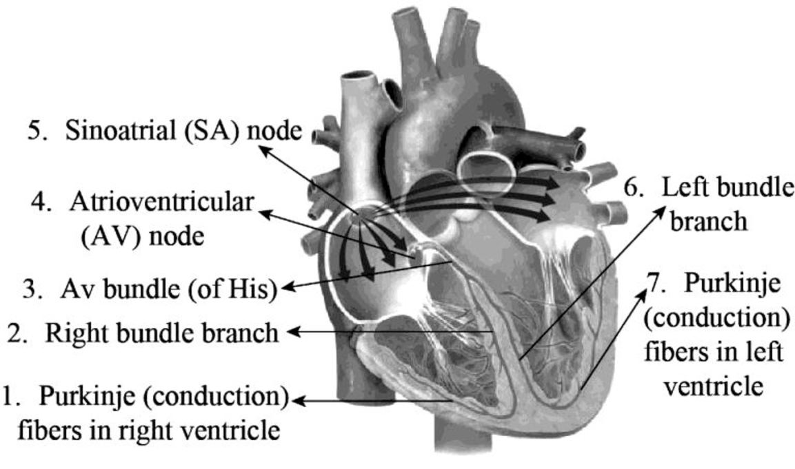
Fig.1: The structures of the cardiac conduction system
Explanation of Solution
The major components of the cardiac conduction system are the SA node, Purkinje fibers, AV node, bundle branches (left and right), and the bundle of His.
Purkinje or conduction fibers are found in the base of the myocardium and in the lateral walls of both the ventricles. They provide the ventricular muscle fibers of the heart and papillary muscles with action potential and induce them to contract.
The left and the right bundle branches are embedded in the interventricular septum and transmit action potential to the conduction fibers.
The AV bundle otherwise known as the bundle of His is located between the atria and the ventricles in a membranous septum that lies superior to the interventricular septum. It is the electrical link amid the ventricles and atria that transmits the action potential to the bundle branches.
The muscle fibers of atria provide action potential for the atrioventricular (AV) node that is present in the lower part of the interatrial septum. It lies in the front part of the coronary sinus opening and supplies action potential to the AV (bundle of His).
The sinoatrial (SA) node is otherwise known as the sinus node. It acts as a natural pacemaker of the heart. This node consists of a group of cells that produce electrical impulses. These impulses pass through the atrial walls and make them contract.
Want to see more full solutions like this?
Chapter 28 Solutions
Laboratory Manual for Anatomy and Physiology, 6e Loose-Leaf Print Companion


 Human Anatomy & Physiology (11th Edition)BiologyISBN:9780134580999Author:Elaine N. Marieb, Katja N. HoehnPublisher:PEARSON
 Biology 2eBiologyISBN:9781947172517Author:Matthew Douglas, Jung Choi, Mary Ann ClarkPublisher:OpenStax
 Anatomy & PhysiologyBiologyISBN:9781259398629Author:McKinley, Michael P., O'loughlin, Valerie Dean, Bidle, Theresa StouterPublisher:Mcgraw Hill Education,
 Molecular Biology of the Cell (Sixth Edition)BiologyISBN:9780815344322Author:Bruce Alberts, Alexander D. Johnson, Julian Lewis, David Morgan, Martin Raff, Keith Roberts, Peter WalterPublisher:W. W. Norton & Company
 Laboratory Manual For Human Anatomy & PhysiologyBiologyISBN:9781260159363Author:Martin, Terry R., Prentice-craver, CynthiaPublisher:McGraw-Hill Publishing Co.
 Inquiry Into Life (16th Edition)BiologyISBN:9781260231700Author:Sylvia S. Mader, Michael WindelspechtPublisher:McGraw Hill Education