
Laboratory Manual For Human Anatomy & Physiology
4th Edition
ISBN: 9781260159080
Author: Martin, Terry R., Prentice-craver, Cynthia
Publisher: Mcgraw-hill Education,
expand_more
expand_more
format_list_bulleted
Concept explainers
Videos
Textbook Question
Chapter 37, Problem F37.11A
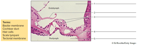
Label the structures indicated in the micrograph of the spiral organ in figure 37.11.
FIGURE 37.11 Label me structures associated with this spiral organ region of a cochlea, using the terms provided (400x)
Expert Solution & Answer
Want to see the full answer?
Check out a sample textbook solution
Students have asked these similar questions
Label the gustatory structures in Figure 24.18(a), (b), and (c).
Examining Microscopic Structure of Cochlea – Organ of Corti (spiral organ) Draw
Draw, Color & Label:
Basilar membrane,
vestibular membrane,
hair cells,
cochlear nerve (afferent fibers),
scala vestibuli,
scala tympani
cochlear duct
organ of corti
Label the structures of the olfactory epithelium in Figure (a) and (b).
Chapter 37 Solutions
Laboratory Manual For Human Anatomy & Physiology
Ch. 37 - Hearing is interpreted in the lobe of the...Ch. 37 - Sound loudness is measured in a. pitch. b....Ch. 37 - The middle ear bones articulate from tympanic...Ch. 37 - The test is done to assess possible conduction...Ch. 37 - Which of the following structures is part of the...Ch. 37 - The pharyngotympanic tube connects the outer e to...Ch. 37 - The cochlear nerve serves as the hearing branch of...Ch. 37 - Endolymph is located within the cochlear duct of...Ch. 37 - FIGURE 37.9 Label the structures associated with...Ch. 37 - FIGURE 37.10 Identify the features indicated on...
Knowledge Booster


Learn more about
Need a deep-dive on the concept behind this application? Look no further. Learn more about this topic, biology and related others by exploring similar questions and additional content below.Similar questions
- Identify the following labels of theretinaarrow_forwardLabel the following numbers: 31, 32 & 37 of the inner ear (B) and cochlea in transverse position (C)arrow_forwardCorrectly identify the following structures of the cochlea. Cochlear duct Cochlear nerve Scala tympani Scala vestibuli Vestibular membrane Spiral organ Spiral ganglion Oval window Aarrow_forward
- Hearing loss is due to problems with ear canals, ear drum or middle ear and its little bones which are malleus, incus, and stapes. Suggest TWO (2) possible treatments that could be conducted.arrow_forwardCan you help me the name of the label of all the number 1,a, 2, 3, 4, 8, B, of the cochlear model?arrow_forwardFigure 1 shows the internal structure of the ear. Label and Describe in details how A-G structure works collectively in the hearing mechanism.arrow_forward
- You have been infected by a virus that led to an inflammation of your cochlea and the destruction of inner ear hair cells. This situation would result in _____________________. Sensorineural hearing impairment Increased movement of stereocilia towards the kinocilium Central hearing impairment Conductive hearing impairment Loss of endolymph in the cochlear duct Please consider providing a detailed and elaborate explanation of both correct options and incorrect options.arrow_forwardStarting with the auricle, trace a sound wave into the innerear to the point at which action potentials are generatedin the cochlear nerve.arrow_forwardOne of the mechanisms that the auditory system uses to localize sound is referred to as interaural time difference (ITD). Where in the brain does this takes place and explain how this mechanism would enable you to localize a sound coming from your left side.arrow_forward
- Image after (at the back) Retina Short distance Object Object https://ophysics.com/16.html Retina Human Eye Lens The above picture shows the defect of a human eye, where the image of the long distance object is formed before(in- front) the retina: 1) What type of eye defect is shown in the picture? 2) The reason for above eye defect is and 3) How can we correct the above eye defect?arrow_forwardLabel the following on the diagram on p. 1 of this handout and the models in the lab. Ampulla (pl. ampullae) How many do you see on the models? Cochlea Oval window Round window Semicircular canals (ducts) How many do you see on the models? Vestibulocochlear nerve (VIII) Which organ of the inner is responsible for each of the following sensations? Hearing a noise: Your body position when you are lying still on your back:arrow_forwardWhich of the following CORRECTLY contrasts the stereocilia in the cochlea versus the stereocilia in the utricle? a). The stereocilia in the cochlea are surrounded by perilymph, whereas the stereocilia in the utricle are surrounded by endolymph. b). The stereocilia in the cochlea are surrounded by endolymph, whereas the stereocilia in the utricle are surrounded by perilymph. c). The stereocilia in the cochlea are embedded in a tectorial membrane whereas the utricle are embedded in a cupula.d). The stereocilia in the cochlea are embedded in a tectorial membrane whersas the stereocilia in the embedded in a cupula.arrow_forward
arrow_back_ios
SEE MORE QUESTIONS
arrow_forward_ios
Recommended textbooks for you
 Understanding Health Insurance: A Guide to Billin...Health & NutritionISBN:9781337679480Author:GREENPublisher:Cengage
 Anatomy & PhysiologyBiologyISBN:9781938168130Author:Kelly A. Young, James A. Wise, Peter DeSaix, Dean H. Kruse, Brandon Poe, Eddie Johnson, Jody E. Johnson, Oksana Korol, J. Gordon Betts, Mark WomblePublisher:OpenStax College
Understanding Health Insurance: A Guide to Billin...
Health & Nutrition
ISBN:9781337679480
Author:GREEN
Publisher:Cengage
Anatomy & Physiology
Biology
ISBN:9781938168130
Author:Kelly A. Young, James A. Wise, Peter DeSaix, Dean H. Kruse, Brandon Poe, Eddie Johnson, Jody E. Johnson, Oksana Korol, J. Gordon Betts, Mark Womble
Publisher:OpenStax College
The Cell Membrane; Author: The Organic Chemistry Tutor;https://www.youtube.com/watch?v=AsffT7XIXbA;License: Standard youtube license