
Concept explainers
Videos
SCIENTIFIC THINKING Microtubules often produce movement through their interaction with motor proteins. But in some cases, microtubules move cell components when the length of the microtubule changes. Through a series of experiments, researchers determined that microtubules grow and shorten as tubulin proteins are added or removed from their ends. Other experiments showed that microtubules make up the spindle apparatus that “pulls” chromosomes toward opposite ends (poles) of a dividing cell. The figures below describe a clever experiment done in 1987 to determine whether a spindle microtubule shortens (depolymerizes) at the end holding a chromosome or at the pole end of a dividing cell.
Experimenters labeled the microtubules of a dividing cell from a pig kidney with a yellow fluorescent dye. As shown on the left half of the diagram below, they then marked a region halfway along the microtubules by using a laser to eliminate the fluorescence from that region. They did not mark the other side of the spindle (right side of the figure).
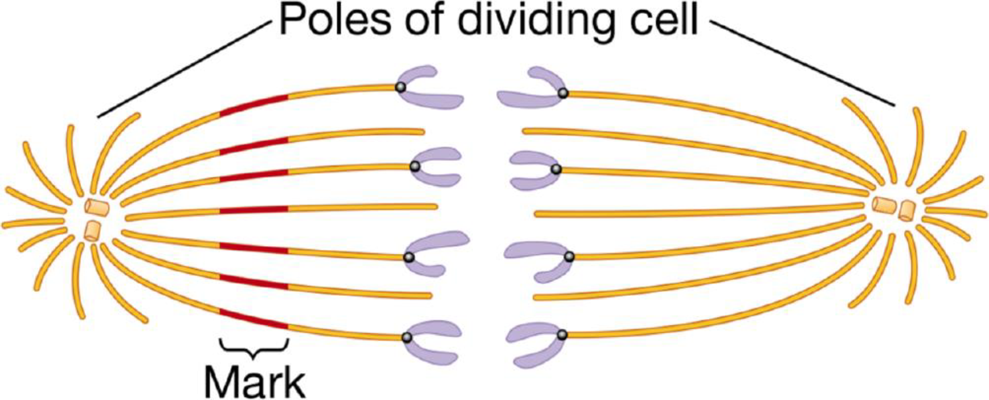
The figure below illustrates the results they observed as the chromosomes moved toward the opposite poles of the cell.
The figure below illustrates the results they observed as the chromosomes moved toward the opposite poles of the cell.
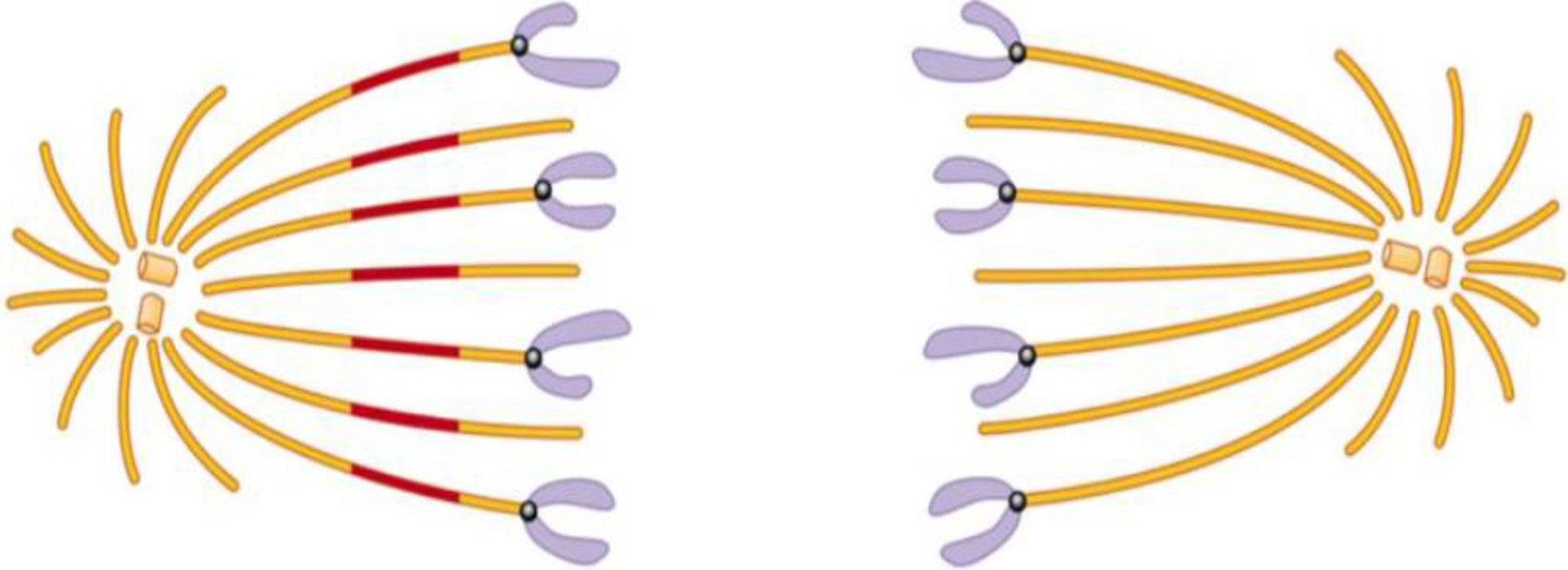
Describe these results. What would you conclude about where the microtubules depolymerize from comparing the length of the microtubules on either side of the mark? How could the experimenters determine whether this is the mechanism of chromosome movement in all cells?
Want to see the full answer?
Check out a sample textbook solution
Chapter 4 Solutions
CAMPBELL BIO&VALPK A/C MAST PKG


- As a cell grows, what happens to its surface area to volume ratio? (Hint: Think of a balloon being blown up). How does this ratio change with respect to cell division?arrow_forwardFrom the image, what is the normal function of the missing structure? A. A lipid bilayer controlling the movement of substances into and out of the cell B. A motor protein that "crawls" along microtubules to contract and release them, which provides movement C. A cytoskeleton element composed of tubulin subunits to provide structure for movement of the cellarrow_forwardcertain cells in the pancreas of animals produce and secrete insulin. Which of the following organelles would be found in abundance in these cells? a.) centrioles and spindle fibers b.) lysosomes and peroxisomes c.) rough ER and golgi apperatusarrow_forward
- As a cell grows, what happens to its surface area to volume ratio? (Hint: Think of a balloon being blown up). How does this ratio change with respect to cell division? Answer in the simplest form.arrow_forwardwhat subunits are microtubules composed of? a. actin b. amino acids c. tubulin d. glucose e. nucleotidesarrow_forwardPart of cell division is that chromosomes are moved the center of the cell and then back to the poles of each newly made daughter cell. These chromosomes are moved to the center of the cell by.. Select one: a. Myosin walking on microtubules towards the center of the cell (slow growing end of microtubule) b. Oar-like motion of myosin with microfilaments causing each half of the cell to move closer together c. kinesins walking on microtubules towards the center of the cell (fast growing end of microtubule) d. vesicles floating on microfilaments of the cortical cytoskeletonarrow_forward
- Which are true of centrioles A- They help arrange the contents of animal cells B- they have a part in cell division C- they are not found in higher plant cellsarrow_forwardYou discover a substance that prevents the motor protein in a centromere from attaching to a spindle fiber. What would be a consequence of adding this substance to a cell during mitosis? Group of answer choices A) the chromatin would not coil up to form condensed chromosomes B) the nuclear membrane would not break down C) sister chromatids would not form D) sister chromatids would fail to separatearrow_forwardCount 100 cells and tally them. Remember the phases you learned in Bio 31 and Anatomy? Draw a pie chart graph showing the phases of Allium root tip. Label each of the phases with the % for each phase: prophase metaphase anaphase telophase interphase provide direction of the phase progression--direction in which to read the graph so the phases of mitosis are in proper order.arrow_forward
- Imagine a cell entered mitosis but was unable to make microtubules. At what stage of mitosis would it remain?arrow_forwardAt prophase, a cell has____________chromosomes, ______________chromatids,and ________________molecules of DNAarrow_forwardGeneral biology 1 Draw two cells under high magnification in the space below and label the cell wall, nucleus and cytopoplasm. Draw a stained onion epidermal cells Describe the difference between the stained and unstained cells? What organelles were seen in the Elodea that are not found in the onion skin? Why would you not expect them to be in the onion?arrow_forward
 Biology: The Dynamic Science (MindTap Course List)BiologyISBN:9781305389892Author:Peter J. Russell, Paul E. Hertz, Beverly McMillanPublisher:Cengage Learning