
Laboratory Manual for Human Anatomy & Physiology (Cat Version)
4th Edition
ISBN: 9781259864612
Author: Martin
Publisher: MCGRAW-HILL HIGHER EDUCATION
expand_more
expand_more
format_list_bulleted
Concept explainers
Textbook Question
Chapter 41, Problem F41.7A
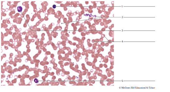
Identify the blood cells indicated in figure 41.7
FIGURE 41.7 Label the specific blood cells on this micrograph of a stained blood smear (400x).
Expert Solution & Answer
Want to see the full answer?
Check out a sample textbook solution
Students have asked these similar questions
I want to do a manual white blood cell differential count on this photo. I just need help with counting how many of each type of WBC there are (example: Lymphocyes-15, Segmented Neutrophils-12, monocytes-7, basophils-3, etc.) **** please note the smudged cells don't need to be counted! Just skip those pls
Label Structure and O2 level
A-
B-
C-
D-
E-
F-
G-
H-
I-
J-
K-
L-
M-
N-
O-
P-
Q-
R-_____ oxygenated blood
S-_____ oxygenated blood
T-
U-
V-
I will make your life a bit easier and not make you count four additional grids. So let's assume this
was a random, relatively uniform sample and arbitrarily assign the other four squares to the exact
same number of red blood cells.
What is the Total red blood cell count? Calculate it as
(#RBC you counted *5)*10,000
Is you answer normal?
Edit View
Insert
Format
Tools
Table
Paragraph v
| в I U
12pt v
Chapter 41 Solutions
Laboratory Manual for Human Anatomy & Physiology (Cat Version)
Ch. 41 - Prob. 1PLCh. 41 - Which of the following is among the agranulocytes?...Ch. 41 - Prob. 3PLCh. 41 - Prob. 4PLCh. 41 - Prob. 5PLCh. 41 - Prob. 6PLCh. 41 - Prob. 7PLCh. 41 - Erythrocytes we also called granulocytes because...Ch. 41 - Complete the following statements: Red blood cells...Ch. 41 - Prob. 2.2A
Ch. 41 - Prob. 2.3ACh. 41 - Prob. 2.4ACh. 41 - Complete the following statements: A mature red...Ch. 41 - Prob. 2.6ACh. 41 - Prob. 2.7ACh. 41 - Prob. 2.8ACh. 41 - Prob. 2.9ACh. 41 - Prob. 2.10ACh. 41 - Complete the following statements: White blood...Ch. 41 - Prob. 2.12ACh. 41 - Prob. 2.13ACh. 41 - Prob. 2.14ACh. 41 - Complete the following statements: Small cell...Ch. 41 - Identify the blood cells indicated in figure 41.7...Ch. 41 - Which leukocyte type would likely be elevated in a...
Additional Science Textbook Solutions
Find more solutions based on key concepts
Sea turtles have disappeared from many regions, and one way of trying to save them is to reintroduce them to ar...
Marine Biology (Botany, Zoology, Ecology and Evolution)
Describe Mendels conclusions about how traits are passed from generation to generation.
Concepts of Genetics (12th Edition)
How does trandlation differ from transcription?
Microbiology: Principles and Explorations
Propose a model for the assembly of a flagellum in a typical Gram-positive cell envelope.
Prescott's Microbiology
Describe Mendels conclusions about how traits are passed from generation to generation.
Concepts of Genetics (11th Edition)
Knowledge Booster


Learn more about
Need a deep-dive on the concept behind this application? Look no further. Learn more about this topic, biology and related others by exploring similar questions and additional content below.Similar questions
- please fill in the charts as the directions states i have included the 2 pictures you need to look at in order Arrange the key and blood smear side by side so that both are visible Use the tally charts below to record the counts for your patient as you go. To count: Start in the upper left corner of the first photo. Identify the white blood cell in the first square and put a mark by its corresponding name on the tally chart. Repeat this process as you move across to the next square and then the next square and so forth. If a square has two WBCs, record both types. There are two sheets of cells to count. Once you have counted all the squares you should have a total of 100 cells. Total the tally for each cell type. Add your total tally for each cell type together to be sure you have a total of 100 recorded. Suggestion: Do not go through and count all the neutrophils, then go through and count all the lymphocytes, then go through and count all the monocytes, etc. Go through the…arrow_forwardAre you able to recognize and identify the various formed elements? You will need to do this is a systematic manner, scanning along the image. The standard method is to use a grid, but this is not possible with this resource. Try constructing a simple table with each leukocyte type and then making a mark for each cell type you identify. Attempt to classify at least 50 and perhaps as many as 100 different cells. Based on the percentage of cells that you count, do the numbers represent a normal blood smear or does something appear to be abnormal?arrow_forwardLabel the parts of the drop of blood in the picture.arrow_forward
- Use Figure 37.2 to identify the leukocytes in the micrograph on the left. __ neutrophil ___ lymphocyte __ eosinophil ____ basophilarrow_forwardMy answer is incorrect, i want to use this to study can you please provide me with the correct answer.arrow_forwardWatch the linked video then answer the questions below. https://youtu.be/HQWlcSp9Sls 1) How many different blood types are there? What are they? 2) What is in the buffy coat? 3) How many different types of solutes are in the blood?arrow_forward
- Label the specific blood cells.arrow_forwardCite one blood disorder following the condition as stated in the first column and discuss each in 2-3 sentences. Name the blood disorder and discuss each in 2-3 sentences. Blood disorder affecting Red Blood Cells Blood disorder affecting White Blood Cells Blood disorder affecting Platelets Blood disorder affecting Blood Plasmaarrow_forwardPlace the following pictures of white blood cells into the appropriate categoryarrow_forward
- Fill in the blank: The shape of a red blood cell can be described as a _______________________ disk.arrow_forwardMicroscope View Slide Blood Description Human blood cells stained with Wright's stain. Red blood cells (erythrocytes) are most numerous and stain pink. White blood cells (leukocytes) are larger with darkly-stained nuclei. Platelets are much smaller than erythrocytes and stain pink. COARSE FOCUS REMOVE SLIDE FINE FOCUS 4X 10X 40X 100X LIGHT ADJUSTarrow_forwardLabel the structures on the pictures, only the listed numbers/letters with the word bank. For this one only numbers listed.arrow_forward
arrow_back_ios
SEE MORE QUESTIONS
arrow_forward_ios
Recommended textbooks for you

 Medical Terminology for Health Professions, Spira...Health & NutritionISBN:9781305634350Author:Ann Ehrlich, Carol L. Schroeder, Laura Ehrlich, Katrina A. SchroederPublisher:Cengage Learning
 Human Biology (MindTap Course List)BiologyISBN:9781305112100Author:Cecie Starr, Beverly McMillanPublisher:Cengage Learning
Medical Terminology for Health Professions, Spira...
Health & Nutrition
ISBN:9781305634350
Author:Ann Ehrlich, Carol L. Schroeder, Laura Ehrlich, Katrina A. Schroeder
Publisher:Cengage Learning
Human Biology (MindTap Course List)
Biology
ISBN:9781305112100
Author:Cecie Starr, Beverly McMillan
Publisher:Cengage Learning