
Laboratory Manual For Human Anatomy & Physiology
4th Edition
ISBN: 9781260159080
Author: Martin, Terry R., Prentice-craver, Cynthia
Publisher: Mcgraw-hill Education,
expand_more
expand_more
format_list_bulleted
Concept explainers
Videos
Textbook Question
Chapter 45, Problem F45.7A
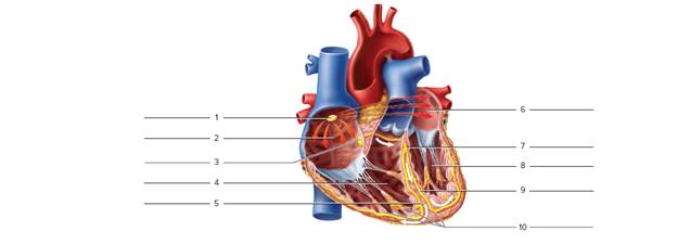
Identify the heart chambers and conduction system structures in the frontal section of the heart in figure 45.7.
FIGURE 45.7 Label the indicated heart chambers and conduction system structures (anterior view).
Expert Solution & Answer
Want to see the full answer?
Check out a sample textbook solution
Students have asked these similar questions
NOTES
and the left side serving the systemic circuit. In Figure 16-15, complete
the schematic showing the blood flow to and from the heart (the starting
points are given to you). Use a blue pen or pencil to denote the direction of
deoxygenated blood and a red pen or pencil for oxygenated blood flow.
Include the names of the major vessels, chambers, and valves involved,
based on the following list:
lung capillary beds
body capillary beds
right ventricle
left ventricle
bicuspid valve
superior vena cava
tricuspid valve
inferior vena cava
pulmonary semilunar valve
pulmonary trunk
aortic semilunar valve
R. and L. pulmonary arteries
R. and L. pulmonary veins
aorta
Pulmonary Circulation
Systemic Circulation
Right atrium
Left atrium
URA--YCK
HAT
Lungs
Body
Figure 16-15. Schematic of circulation
Label the veins in Figure
The heart is lateral to the lungs. True or false
Chapter 45 Solutions
Laboratory Manual For Human Anatomy & Physiology
Ch. 45 - The _______ of the conduction system is known as...Ch. 45 - The ________ of the conduction system is/are...Ch. 45 - The first of two heart sounds (lubb) occurs when...Ch. 45 - One cardiac cycle would consist of a. left chamber...Ch. 45 - The SA node of the heart is located in the a....Ch. 45 - The depolarization of ventricular fibers is...Ch. 45 - The dupp sound occurs when the semilunar valves...Ch. 45 - The P wave of an ECG occurs during the...Ch. 45 - The period during a heart is contracting is called...Ch. 45 - The period during which a heart chamber is...
Ch. 45 - During ventricular contraction, the AV valves...Ch. 45 - During ventricular relaxation, the AV valves are...Ch. 45 - The pulmonary and aortic valves open when the...Ch. 45 - The first sound of a cardiac cycle occurs when the...Ch. 45 - The second sound of a cardiac cycle occurs when...Ch. 45 - The sound created when blood leaks back through an...Ch. 45 - What changes did you note in the heart sounds when...Ch. 45 - What changes did you note in the heart sounds...Ch. 45 - Prob. 3.1ACh. 45 - The ____________________ node is located in the...Ch. 45 - The fibers that carry cardiac impulses from the...Ch. 45 - Prob. 3.4ACh. 45 - The P wave corresponds to depolarization of the...Ch. 45 - The QRS complex corresponds to depolarization of...Ch. 45 - The T wave corresponds to repolarization of the...Ch. 45 - Why is atrial repolarization not observed in the...Ch. 45 - Identify the heart chambers and conduction system...Ch. 45 - How much time passed from the beginning of the P...Ch. 45 - What is the significance of this P-R interval?Ch. 45 - How can you determine heart rate from an...Ch. 45 - As blood in the ventricles surges back against the...
Knowledge Booster


Learn more about
Need a deep-dive on the concept behind this application? Look no further. Learn more about this topic, biology and related others by exploring similar questions and additional content below.Similar questions
- ANTERIOR SIDE On the figure seen, the valve marked with the arrow indicates the tricuspid valve bicuspid O pulmonary semilunar valve aortic semilunar valvearrow_forwardPlace the initials for each chamber over the Xs on the heart and then drag each structure to its correct arrow. aortic valve RA pulmonary valve RV tricuspid valve LV bicuspid valvearrow_forwardLabel the dorsal external features of the frog’s heart. Answer all labels on the space provided.arrow_forward
- Place the initials for each chamber over the Xs on the heart and then drag each blood vessel to its correct arrow. aorta RV superior vena cava LV fossa ovalis RA coronary sinus tricuspid valve left pulmonary artery pulmonary valve pulmonary trunkarrow_forwardPlease help me label this, THANK YOUarrow_forwardPlace the initials for each chamber over the Xs on the heart and then drag each blood vessel to its correct arrow. right pulmonary veins LV superior vena cava LA left pulmonary artery X RV right pulmonary artery left pulmonary veins aorta coronary sinus inferior vena cavaarrow_forward
arrow_back_ios
SEE MORE QUESTIONS
arrow_forward_ios
Recommended textbooks for you

 Fundamentals of Sectional Anatomy: An Imaging App...BiologyISBN:9781133960867Author:Denise L. LazoPublisher:Cengage Learning
 Medical Terminology for Health Professions, Spira...Health & NutritionISBN:9781305634350Author:Ann Ehrlich, Carol L. Schroeder, Laura Ehrlich, Katrina A. SchroederPublisher:Cengage Learning
Fundamentals of Sectional Anatomy: An Imaging App...
Biology
ISBN:9781133960867
Author:Denise L. Lazo
Publisher:Cengage Learning
Medical Terminology for Health Professions, Spira...
Health & Nutrition
ISBN:9781305634350
Author:Ann Ehrlich, Carol L. Schroeder, Laura Ehrlich, Katrina A. Schroeder
Publisher:Cengage Learning
Dissection Basics | Types and Tools; Author: BlueLink: University of Michigan Anatomy;https://www.youtube.com/watch?v=-_B17pTmzto;License: Standard youtube license