
Campbell Biology Custom Stony Brook 10 Th Edition
10th Edition
ISBN: 9781269870818
Author: Reece Urry Cain Wasserman Minorsky Jackson
Publisher: PEARSON
expand_more
expand_more
format_list_bulleted
Concept explainers
Videos
Textbook Question
Chapter 47, Problem 8TYU
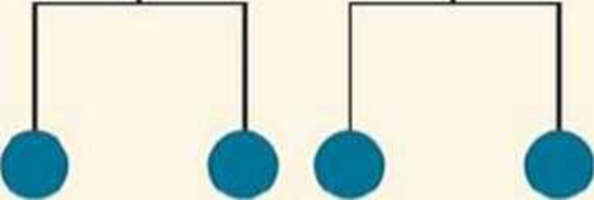
DRAW IT Each blue circle in the figure below represents a cell in a cell lineage. Draw two modified versions of the cell lineage so that each Version produces three cells. Use apoptosis in one of the versions, marking any dead cells with an X.

Expert Solution & Answer
Want to see the full answer?
Check out a sample textbook solution
Students have asked these similar questions
DRAW IT Each blue circle in the figure below represents a cellin a cell lineage. Draw two modified versions of the cell lineageso that each version produces three cells. Use apoptosis in oneof the versions, marking any dead cells with an X.
the light micrograph shows dividing cells near the tip of an onion root. identify and encircle a cell in each of the following stages: phrophase , prometaphase, metaphase ,anaphase and telophase. describe the major events occuring at each stage.
The cell in image is ...
in interphase
S phase
mitosis
G1 phase
and one conclusion that can be drawn is :
TRAP150 co-localizes with DNA
TRAP 150 is cytoplasmic
a clear conclusion cannot be drawn
TRAP 150 does not co localize with dna
Chapter 47 Solutions
Campbell Biology Custom Stony Brook 10 Th Edition
Ch. 47.1 - How does the fertilization envelope form in sea...Ch. 47.1 - Prob. 2CCCh. 47.1 - MAKE CONNECTIONS Review Figure 12.16 on cell...Ch. 47.2 - In the frog embryo, convergent extension elongates...Ch. 47.2 - WHAT IF? Predict what would happen if Ca2+ was...Ch. 47.2 - MAKE CONNECTIONS Unlike some other types of birth...Ch. 47.3 - Prob. 1CCCh. 47.3 - Prob. 2CCCh. 47.3 - Prob. 3CCCh. 47.3 - Prob. 4CC
Ch. 47 - What cell-surface event would likely fail if a...Ch. 47 - Prob. 47.2CRCh. 47 - Suppose you found two classes of mouse mutations,...Ch. 47 - Prob. 1TYUCh. 47 - Which of the following is common to the...Ch. 47 - The archenteron develops into a. the mesoderm. b....Ch. 47 - What structural adaptation in chickens allows them...Ch. 47 - Prob. 5TYUCh. 47 - In humans, identical twins are possible because a....Ch. 47 - Cells transplanted from the neural tube of a frog...Ch. 47 - DRAW IT Each blue circle in the figure below...Ch. 47 - EVOLUTION CONNECTION Evolution in insects and...Ch. 47 - Prob. 10TYUCh. 47 - Prob. 11TYUCh. 47 - Prob. 12TYUCh. 47 - SYNTHESIZE YOUR KNOWLEDGE Occasionally, two-headed...
Knowledge Booster


Learn more about
Need a deep-dive on the concept behind this application? Look no further. Learn more about this topic, biology and related others by exploring similar questions and additional content below.Similar questions
- Review the image below. What cell division process is the image demonstrating? Provide evidence to support your claim. * In your response, you should explain what type and kind of cells are being produced? You should compare the chromosomes in the parent cell and daughter cells. Response should be at least 3 SENTENCES (I) S-phase Two daughter cells DNA replication Parent cell Your answer hoct describes a human with the chromosomes represented in thearrow_forwardDraw and label a picture of what a cell looks like during prophase. Draw and label a picture of what a cell looks like during metaphase. Draw and label a picture of what a cell looks like during anaphase. Draw and label a picture of what a cell looks like during telophase. Draw a picture of what a cell looks like during cytokinesis. Draw and label a picture of prophase I & II. Draw and label a picture of metaphase I & II.arrow_forwardCleavage furrow formed during cytokinesis is found in? A-both in animal and plants B- neither in animal nor in plant cells C- plant cells D- animal cellsarrow_forward
- The genes below have been knocked out (loss of function). Draw what the cell would look like during the appropriately affected stage of mitosis. State what stage you are depicting on your drawing. (Each gene knockout is occurring in a different cell; you should have a drawing of the affected cell for each). 1. Separase, 2. Cohesinarrow_forwardThe following diagram shows a simulated microscopic view of a tissue sample taken from a patient where the cells have been squashed onto a slide and stained to visualize the cell's DNA. In the diagram, click or tap on the center of all cells that appear to be involved in any of the four stages of mitosis. Make sure to mark as close to the center of the cell as possible. In the diagram from Question 5, count the total number of cells visible and write the total number of cells in the space below: In the diagram from Question 5, count the number of cells that you had marked as currently undergoing any of the four phases of mitosis. How many cells in total are currently undergoing mitosis? Write this number in the space below: The mitotic index is a calculated value that represents the percentage of cells in a sample that are actively dividing. It involves counting the total number of cells present including those actively dividing or those in interphase, the total number of cells that…arrow_forwardChoose all of the TRUE statements. Hint: 4 statements are true. Cytokinesis occurs the same way in both plant and animal cells. Sister chromatids are genetically identical. The term chromosome can be used to describe both chromosomes composed of one and two chromatids. Mitosis results in daughter cells that are genetically identical to the parent cell. All eukaryotic somatic cells are diploid. Cyclin dependent kinases are present but not active in cell cycle regulation without the presence of cyclin proteins. Chromosomes are always visible in the cell.arrow_forward
- The figure below shows the number of chromosomes observed in an actively dividing cell at each stage of cell division. A B number of chromsomes per cell C 100 90 D 80 A bar graph comparing the number of chromosomes at different stages of cell division. 20 Which of the following best explains the change in the number of chromosomes between metaphase and anaphase? prophase metaphase anaphase telophase cytokinesis stage of cell division New chromosomes formed during prophase are doubled during anaphase. DNA replication occurs between metaphase and anaphase, doubling the number of chromosomes. During metaphase, a cell contains identical copies of each chromosome, and then trans- forms into sister chromatids. During anaphase, the chromatids are separated, each becoming independent chromo- somes in its respective new cellarrow_forwardDrag the terms on the left to the appropriate blanks on the right to complete the sentences. Reset Help breaks in chromosomes The two resulting daughter cells completely identical if divide first and then duplicate advantageous chromosomes. The replication that occurs after division would generate which would be be repaired according to the cell. In this case, the genetic information inherited less accurately. This way of cell division would be parent for organisms and would likely be daughter mutations spread would not be deletions eliminated couldn't could disadvantageous P Pearsonarrow_forwardv Part A Match the description with the stage of the cell cycle. Reset Help Centrioles move to opposite ends of the cell DNA condenses to form chromosomes Chromosomes line up in the middle of the cell Chromosomes Split and Move to opposite ends of the cell Cell begins to split into two DNA replicates Interphase Prophase Metaphase Anaphase Telophase UnitConversionSE.pdf Type here to search 99+ RB (1arrow_forward
- What choice best describes what happens immediately following the image below in the process of cell division. (level 3) The cytoplasms splits Sister chromatids are seperated to opposite sides of the cell chromatin condenses into chromatids a new nuclear envelope is formed stv MacBook Air DII F10 80 F4 F3 & #3 2$ 7 8. 9 3 4 5 9. P Y W E R F G H J C V command nd .. .- レ V * 00arrow_forwardThis is a picture of anaphase: Explain why you know this cell is in anaphase using 2 vocabulary words from this list (definitions are in question 1): Nuclear envelope/nucleus Chromatin chromatid/sister chromatid Chromosome Spindle tv Aa 23 MacBook Air DD FB F9 F7 F3 F4 F5 F2 @ 23 $ % & 3 4 5 7 8 9 P W R Y S D F G H J K L XIG YIB N M Earrow_forwardattached image is an image of the cells you would have been looking at under the microscope Count at least 50 cells. When done, there should be at least 50 marks on the table (one mark for each cell). You may need to count more than 50 to find at least one of every phase. Calculate the proportions, and estimate the amount of time spent in each phase and subphase. The table will lead you through how this works. Take the number of cells in a particular phase, divided by the total number of cells examined (i.e., 50—or more), then multiply by 24 (the number of hours an average onion root tip cell takes to complete the entire cycle. This should give the hours a cell spends in each phase. Again, this assumes, that the entire cell cycle for onion root cells is 24 hours. This time can vary in different organisms. FILL-IN this table with the results of your count of 50 cells Phase/Subphase # of cells Calculation (fraction of cells x total hours of the cell cycle) # hours spent…arrow_forward
arrow_back_ios
SEE MORE QUESTIONS
arrow_forward_ios
Recommended textbooks for you
 Biology (MindTap Course List)BiologyISBN:9781337392938Author:Eldra Solomon, Charles Martin, Diana W. Martin, Linda R. BergPublisher:Cengage Learning
 Biology Today and Tomorrow without Physiology (Mi...BiologyISBN:9781305117396Author:Cecie Starr, Christine Evers, Lisa StarrPublisher:Cengage Learning
 Biology 2eBiologyISBN:9781947172517Author:Matthew Douglas, Jung Choi, Mary Ann ClarkPublisher:OpenStax
 Human Biology (MindTap Course List)BiologyISBN:9781305112100Author:Cecie Starr, Beverly McMillanPublisher:Cengage Learning
Biology (MindTap Course List)
Biology
ISBN:9781337392938
Author:Eldra Solomon, Charles Martin, Diana W. Martin, Linda R. Berg
Publisher:Cengage Learning
Biology Today and Tomorrow without Physiology (Mi...
Biology
ISBN:9781305117396
Author:Cecie Starr, Christine Evers, Lisa Starr
Publisher:Cengage Learning
Biology 2e
Biology
ISBN:9781947172517
Author:Matthew Douglas, Jung Choi, Mary Ann Clark
Publisher:OpenStax
Human Biology (MindTap Course List)
Biology
ISBN:9781305112100
Author:Cecie Starr, Beverly McMillan
Publisher:Cengage Learning
The Cell Cycle and its Regulation; Author: Professor Dave Explains;https://www.youtube.com/watch?v=eqJqhA8HSJ0;License: Standard YouTube License, CC-BY
Cell Division - Mitosis and Meiosis - GCSE Biology (9-1); Author: Mr Exham Biology;https://www.youtube.com/watch?v=w7vp_uRA8kw;License: Standard YouTube License, CC-BY