
LAB MANUAL FOR HUMAN A&P MAIN VERSION
4th Edition
ISBN: 9781266871016
Author: Martin
Publisher: MCG
expand_more
expand_more
format_list_bulleted
Videos
Textbook Question
Chapter 47, Problem F47.17A
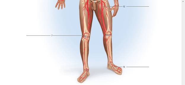
Label the major arteries and veins indicated in figures 47.17 and 47.18.
FIGURE 47.17 Label the major systemic arteries.

Expert Solution & Answer
Want to see the full answer?
Check out a sample textbook solution
Students have asked these similar questions
Label the veins in Figure
Identify the layers of this artery labelled “A” and “B”
ANTERIOR SIDE
On the figure seen, the valve marked with the arrow indicates the
tricuspid valve
bicuspid
O pulmonary semilunar valve
aortic semilunar valve
Chapter 47 Solutions
LAB MANUAL FOR HUMAN A&P MAIN VERSION
Ch. 47 - The middle layer of an artery and vein contains...Ch. 47 - Arteries carry blood a. away from the heart. b....Ch. 47 - The ________ has the thickest wall. a. right...Ch. 47 - Which of the following arteries is not a direct...Ch. 47 - Which of the following veins is nor located in the...Ch. 47 - Which of the following arteries is part of the...Ch. 47 - A capillary wall is composed of three tunic s...Ch. 47 - The left ventricle pumps blood into the aorta of...Ch. 47 - Sketch and label a section of an arterial wall...Ch. 47 - Describe the differences you noted in the...
Ch. 47 - Describe the differences you noted in the...Ch. 47 - How did you distinguish between arterioles and...Ch. 47 - How did you recognize capillaries in the web?Ch. 47 - Brachiocephalic trunk, _____, right axillary...Ch. 47 - Ascending aorta, ______, descending thoracic aortaCh. 47 - Abdominal aortae _______, ascending colon (right...Ch. 47 - Brachiocephalic trunk,_________ , right external...Ch. 47 - Axillary artery, ______, radial arteryCh. 47 - Common iliac artery, _____, femoral arteryCh. 47 - Pulmonary trunk, _____, lungsCh. 47 - Brachiocephalic trunk, _______, right axillary...Ch. 47 - Ascending aorta ___________, descending thoracic...Ch. 47 - Abdominal aortae ________, ascending colon (right...Ch. 47 - Brachiocephalic trunk, ________, right external...Ch. 47 - Axillary artery, _______, radial arteryCh. 47 - Common iliac artery, _______, femoral arteryCh. 47 - Pulmonary trunk, _____, lungsCh. 47 - Label the major arteries and veins indicated in...Ch. 47 - FIGURE 47.18 Label the major systemic veins.
Knowledge Booster


Learn more about
Need a deep-dive on the concept behind this application? Look no further. Learn more about this topic, biology and related others by exploring similar questions and additional content below.Similar questions
- Identify each artery in Figure and write its name next to the number.arrow_forward_____ artery originates from the ventral wall of the common iliac artery.arrow_forwardNOTES and the left side serving the systemic circuit. In Figure 16-15, complete the schematic showing the blood flow to and from the heart (the starting points are given to you). Use a blue pen or pencil to denote the direction of deoxygenated blood and a red pen or pencil for oxygenated blood flow. Include the names of the major vessels, chambers, and valves involved, based on the following list: lung capillary beds body capillary beds right ventricle left ventricle bicuspid valve superior vena cava tricuspid valve inferior vena cava pulmonary semilunar valve pulmonary trunk aortic semilunar valve R. and L. pulmonary arteries R. and L. pulmonary veins aorta Pulmonary Circulation Systemic Circulation Right atrium Left atrium URA--YCK HAT Lungs Body Figure 16-15. Schematic of circulationarrow_forward
- Please help me identify this diagramarrow_forwardThe red blood cell took the posterior tibial vein between the plantar veins and the femoral vein. What other vein could it have taken instead of the posterior tibial vein? _______________arrow_forwardIdentify each vein in Figure and write its name next to the number.arrow_forward
- labelarrow_forwardPath from median antecubital vein at the left elbow, to the tibial vein on the right leg.arrow_forwardLabel the below figure with the correct artery using the list of arteries provided. Subclavian artery Radial artery Digital arteries Brachial artery Ulnar artery Deep volar (palmar) arch Superficial volar (palmar) arch Axillary artery Glierdon-McNeil, LLC 1. 2. 3. 5. 6. 7. 8.arrow_forward
- The patient was given IV (intravenous) with medication into the left great saphenous vein. Describe flow of the medication through the blood vessels into the right atrium. Specify all blood vessels on the way from the left great saphenous vein into the right atrium.arrow_forwardA pulse can be felt in the following arteries: superficial temporal, facial, common carotid, brachial, femoral, popliteal, posterior tibial, and dorsalis pedis (Figure 20.8b). Name the artery from which each artery listed branches. Here is an example to get you started: The superficial temporal artery is a branch of the external carotid artery.arrow_forwardSignificant ST-segment elevation represents potential myocardialarrow_forward
arrow_back_ios
SEE MORE QUESTIONS
arrow_forward_ios
Recommended textbooks for you

 Fundamentals of Sectional Anatomy: An Imaging App...BiologyISBN:9781133960867Author:Denise L. LazoPublisher:Cengage Learning
 Medical Terminology for Health Professions, Spira...Health & NutritionISBN:9781305634350Author:Ann Ehrlich, Carol L. Schroeder, Laura Ehrlich, Katrina A. SchroederPublisher:Cengage Learning
Fundamentals of Sectional Anatomy: An Imaging App...
Biology
ISBN:9781133960867
Author:Denise L. Lazo
Publisher:Cengage Learning
Medical Terminology for Health Professions, Spira...
Health & Nutrition
ISBN:9781305634350
Author:Ann Ehrlich, Carol L. Schroeder, Laura Ehrlich, Katrina A. Schroeder
Publisher:Cengage Learning
The Cardiovascular System: An Overview; Author: Strong Medicine;https://www.youtube.com/watch?v=Wu18mpI_62s;License: Standard youtube license