Laboratory Manual For Human Anatomy & Physiology
4th Edition
ISBN: 9781260159080
Author: Martin, Terry R., Prentice-craver, Cynthia
Publisher: Mcgraw-hill Education,
expand_more
expand_more
format_list_bulleted
Videos
Textbook Question
Chapter 50, Problem 2.1A
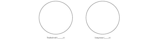
Each circle below represents the microscopic field of view. In each circle, sketch and label a portion of the tracheal wall and a portion of lung tissue.
Expert Solution & Answer
Want to see the full answer?
Check out a sample textbook solution
Students have asked these similar questions
Both arrows must point to the same structure in the trachea. Based on the pseudostratified epithelium. Which
The highlighted structure in the above image is a(n):
Granuloma
Pulmonary artery
Bronchiole
Alveolar sac
Write on a respiratory system (Please include the following point (structure of respiratory system etc)
Chapter 50 Solutions
Laboratory Manual For Human Anatomy & Physiology
Ch. 50 - The right lung has lobes ______ the left lung has...Ch. 50 - Paranasal sinuses are within the following bones...Ch. 50 - The alveoli are composed of a. simple squamous...Ch. 50 - The _________ adheres to the surface of the lung....Ch. 50 - The ________ is the most inferior cartilage of the...Ch. 50 - Prob. 6PLCh. 50 - Which of the following airway tubes would have the...Ch. 50 - Prob. 8PLCh. 50 - Match the terms in column A with the descriptions...Ch. 50 - Label the structures indicated in figure 50.11,...
Ch. 50 - FIGURE 50.12 Label the features of the upper...Ch. 50 - FIGURE 50.13 Label the features of the larynx...Ch. 50 - Each circle below represents the microscopic field...Ch. 50 - What is the function of the mucus secreted by the...Ch. 50 - Describe the function of the cilia in the...Ch. 50 - How is breathing affected if the smooth muscle of...Ch. 50 - How is breathing affected if the smooth muscle of...Ch. 50 - What is the functional advantage of the alveolar...Ch. 50 - What affect would pulmonary edema have on this...Ch. 50 - Describe the airway of a patient who is having an...
Knowledge Booster


Learn more about
Need a deep-dive on the concept behind this application? Look no further. Learn more about this topic, biology and related others by exploring similar questions and additional content below.Similar questions
- Write on a respiratory system (Please include the following points (function of parts of the systems etc)arrow_forwardRespiratory System in Insects (air-dried insect and whole mount of insect spiracle and trachea) Parameter Identification/Description/Enumeration Shape of the spiracle Body Segments where spiracles are found Shape of the tracheaarrow_forwardHow would this action change the volume of the chest and lungs? Why does it cause the person to expel the food item from the airway?Match the words in the left column to the appropriate blanks in the sentences on the right.arrow_forward
- Write on a respiratory system (function of parts of the systems in details)arrow_forwardOn the photomicrograph of lung. Identify tube A bronchiole Identify space B The epithelium lining A is + epithelium. The epithelium lining B is epithelium. Tissue C is | Please answer all parts of the question. B Aarrow_forwardUse the figure to match the following. Drag the appropriate labels to their respective targets. Trachea Carina of trachea Pharynx Main (primary) bronchus Larynx Submit Request Answer Right lung Left lung Diaphragm Reset Helparrow_forward
- Why is the pulmonary artery colored blue in this figure? Answer can be found in Appendix F. Blood flow Blood flow - Venule Arteriole Simple squamous epithelial cells Alveolar wall Capillary - Alveolus Air Alveolus co, co, 0, Figure 16.9 Light micrograph of alveoli (250x). APIR O McGraw-Hill Education/Al Telser, photographer Capillary Figure 16.10 Oxygen (O2) diffuses from air within thearrow_forwardIdentify the parts if the lungs using numbers.arrow_forwardFill in the blank: Only a thin film of lubricating serous fluid separates the parietal pleura from the _______________________ of a lung.arrow_forward
- FIGURE 13.11 Respiratory structures (cont.): (B) the larynx, anterior view; (C) the larynx, midsagittal section B O Arytenoid cartilage O Corniculate cartilage O Cricoid cartilage O Cuneiform cartilage O Epiglottis O Epiglottic cartilage O False vocal cords O Fat pad O Hyoid bone O Thyroid cartilage O Tracheal cartilage O True vocal cords 2 Label Figure 13.12 with the terms below. O Alveolar duct O Alveolar sac O Alveolus O Pulmonary arteriole O Pulmonary capillaries O Pulmonary venule O Respiratory bronchiole O Terminal bronchiolearrow_forward1 Label Figure 13.11A and 13.11B with the terms below. Note that 13.11B is on the next page. O Diaphragm O Laryngopharynx O Larynx O Left primary bronchus O Nasal cavity O Nasopharynx O Oropharynx O Pleural cavity O Right primary bronchus O Secondary bronchi TICURE 13.11 Respiratory structures: (A) the lungs and respiratory tract (continues)arrow_forwardDescribe the histology of the bronchial tree. Emphasize on the epithelium and other types of tissues they specifically have. Include also the structures/cells present in your discussion. Please do not copy and paste from previous answer.arrow_forward
arrow_back_ios
SEE MORE QUESTIONS
arrow_forward_ios
Recommended textbooks for you

 Medical Terminology for Health Professions, Spira...Health & NutritionISBN:9781305634350Author:Ann Ehrlich, Carol L. Schroeder, Laura Ehrlich, Katrina A. SchroederPublisher:Cengage Learning
- Basic Clinical Lab Competencies for Respiratory C...NursingISBN:9781285244662Author:WhitePublisher:Cengage
Medical Terminology for Health Professions, Spira...
Health & Nutrition
ISBN:9781305634350
Author:Ann Ehrlich, Carol L. Schroeder, Laura Ehrlich, Katrina A. Schroeder
Publisher:Cengage Learning
Basic Clinical Lab Competencies for Respiratory C...
Nursing
ISBN:9781285244662
Author:White
Publisher:Cengage
Respiratory System; Author: Amoeba Sisters;https://www.youtube.com/watch?v=v_j-LD2YEqg;License: Standard youtube license