
LABORATORY MANUAL FOR HUMAN ANATOMY & PH
4th Edition
ISBN: 9781260254426
Author: Martin
Publisher: MCG
expand_more
expand_more
format_list_bulleted
Videos
Textbook Question
Chapter 57, Problem F57.1A
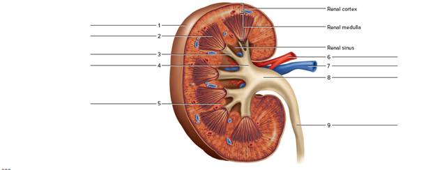
Label the features indicated in figure 57.10 of a kidney (frontal section).
FIGURE 57.10 Label the structures in the frontal section of a kidney.
Expert Solution & Answer
Want to see the full answer?
Check out a sample textbook solution
Students have asked these similar questions
Observe the X-ray in Figure and identify the structures. FIGURE Urinary system structures in the cat.
Observe the X-ray in Figure and identify the structures.
1 Label the following parts of the kidney on Figure 14.14.
O Major calyx
Minor calyx
ORenal artery
Renal capsule
Renal column
Renal cortex
Renal medulla
Renal pelvis
Renal pyramid
Renal vein
O Ureter
Chapter 57 Solutions
LABORATORY MANUAL FOR HUMAN ANATOMY & PH
Ch. 57 - When comparing the position of the two kidneys. a....Ch. 57 - Prob. 2PLCh. 57 - Which of the following does not represent one of...Ch. 57 - The ______________ arteries and veins are located...Ch. 57 - The _____________ is the tube from the kidney to...Ch. 57 - The trigone is a triangular, funnel-like region of...Ch. 57 - The external urethral sphincter is composed of...Ch. 57 - Contractions of the detrusor muscle provide the...Ch. 57 - Label the features indicated in figure 57.10 of a...Ch. 57 - Match the terms in column A with the descriptions...
Knowledge Booster


Learn more about
Need a deep-dive on the concept behind this application? Look no further. Learn more about this topic, biology and related others by exploring similar questions and additional content below.Similar questions
- IDENTIFY THE RENAL PELVIS А- 0 0 0 0 0 A В E B . D Earrow_forwardFUNCTIONS OF THE KIDNEY STRUCTURES Figure 15-5 AB BC AC AD BD BE CD AE CE 27. Which label in Figure 15-5 is described as a collection area of the kidney that is continuous with the ureter? 28. Which label in Figure 15-5 carries unfiltered blood?arrow_forwardLable itarrow_forward
- FIGURE 9-6 The renal corpuscle. immediately after Bowman's capsule. It becomes the loop of Henle (or nephron loop) as it moves deep and forms the distal convoluted tubule away from Bowman's capsule. Distal convoluted tubules from many nephrons join to form a collecting duct that drains urine from the medulla to the renal papillae. Now label Figure 9-7. H TERMS FOR FIGURE 9-7 Afferent arteriole Bowman's capsule Collecting duct Distal convoluted tubule Efferent arteriole Glomerulus Interlobular artery Interlobular vein Loop of Henle Peritubular capillaries Proximal convoluted tubule FIGURE 9-7 The nephron and blood supply. LAB 9 Urinary System 83 processed to become urine. The part. It is called the proximarrow_forwardDraw a damaged kidney with kidney stones. Explain how it occurs including the processes inside the urinary system. ( Please make it clean and readable, labelled should be included).[ Drawing ] Thank youarrow_forwardPlace the following vessels in the order in which blood flows through 1. v glomerulus 2. v renal artery 3. v interlobar artery 4. v arcuate artery 5. v cortical radiate arteryarrow_forward
arrow_back_ios
SEE MORE QUESTIONS
arrow_forward_ios
Recommended textbooks for you

Excretory System; Author: Amoeba Sisters;https://www.youtube.com/watch?v=q5qaGHfdmYM;License: Standard youtube license