
Concept explainers
Videos
The microarray shown at the top of this page analyzes genomic DNA from a nuclear family in which the father, one son, and one daughter have rare, late onset polycystic kidney disease; while the mother, the second son, and the second daughter are unaffected. As stated in previous problem, the eight SNP loci examined are evenly spaced at about 10 Mb intervals on chromosome 4, and they are shown on the microarray in their actual order on this chromosome.
| a. | Is the allele responsible for the disease dominant or recessive with respect to wild type? Is the disease gene autosomal or X-linked? |
| b. | For each of the four siblings, indicate the genotype of the sperm from which they were created. For each of these sperm, write the alleles for each of the eight loci on chromosome 4 in order. |
| c. | Identify the two SNP loci that are uninformative in this family (that is, you cannot determine whether or not either of these loci is linked to the disease gene). |
| d. | Assuming for the sake of simplicity that the four children shown would be completely representative even if the parents had 100 children, the data in the figure indicate that one locus is unlinked to the disease gene. Which one? |
| e. | The microarray results indicate that during meiosis in the father, two different crossovers occurred in the region including the disease gene and the SNP loci that are genetically linked to it. Draw a map of chromosome 4 showing the locations of the disease gene, the linked SNP loci, and the two recombination events. Your map should indicate any uncertainties in these positions. |
| f. | Diagram the location and arrangement (phase) of all alleles of all genes that are on the two chromosomes in the father’s diploid genome. |
a.
To determine:
Whether the allele responsible for the disease is dominant or recessive concerning wild form and whether the disease gene is autosomal or X-linked.
Introduction:
The alleles that reveal their effect even if the individual only has one copy of allele are known as dominant alleles.
Explanation of Solution
The allele that is responsible for the disease is an autosomal dominant allele. Autosome refers to the non-sex chromosome, and the term dominant signifies that a single copy of disease-associated mutation can cause disease.
b.
To determine:
The genotype of the sperm from which the four siblings were formed and for each sperm, order the allele for each of eight loci on chromosome 4.
Introduction:
The male reproductive organs called testes releases sperms. A genotype is the set of genes in the DNA (deoxyribonucleic acid) which causes a particular trait.
Explanation of Solution
| Allele and loci | 1 | 2 | 3 | 4 | 5 | 6 | 7 | 8 |
| Affected father | CA* | GG* | GA* | TG* | CC | GC* | GA* | TG* |
| Unaffected mother | AA | GC | GG | AT | CC | CC | AG | TT |
| Affected son | CA* | CG* | GA* | AG* | CC | CC* | AA* | TT* |
| Affected daughter | AA* | GG* | GA* | TG* | CC | CC* | AA* | TG* |
| Unaffected son | AC | GG | AG | TT | CC | CG | GG | TT |
| Unaffected daughter | AA | CG | GG | TT | CC | CG | GG | TT |
Alleles with asterisk superscript denote disease associated alleles. Alleles shown in red indicate the genotype of sperm from which the siblings were formed.
c.
To determine:
The way to prove that the loci are on chromosome 4 and are 10 Mb apart.
Introduction:
The mating that reveals the inheritance or linkage relationships of a gene or an allele is referred to as informative mating.
Explanation of Solution
For mating to be informative, the loci must be heterozygous in key individuals. If both parents are heterozygous, meiotic events can be easily reconstructed by studying the offspring genotypes. The loci 4 and 7 are heterozygous and are informative, of which allele is inherited from which parent.
d.
To determine:
The locus that is unlinked to the disease gene.
Introduction:
Linked gene refers to the genes whose alleles are mostly inherited together and are these genes are usually present close together on the same chromosome.
Explanation of Solution
Locus 1 is unlinked to disease gene because the father is heterozygous for CA alleles. On the contrary, one of the affected daughters is homozygous for AA alleles, and one who is not affected also has the game genotype.
e.
To draw:
A map of chromosome 4 that show the positions of the disease gene, linked SNP loci, and two crossovers and also indicate any uncertainties in these locations.
Introduction:
The process by which the offspring obtain a combination of alleles different from that of either of the parents is known as recombination.
Explanation of Solution
Pictorial representation: Fig. 1 represents the genetic map of chromosome 4 that indicates the location of the disease gene, linked SNP loci, and two crossovers. Here, A, G, T, and C indicate the SNPs (single nucleotide polymorphisms) at their respective position. The asterisk superscript indicates the genotype of disease alleles.
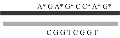
Fig 1. A map of chromosome 4
During meiosis, the recombination occurred at locus 8 between alleles G and T in the father. As a result of recombination, one of the sons inherited recombinant allele. Though the son is homozygous for TT allele, the son became affected due to recombination.
f.
To draw:
The position and arrangement of all alleles of all genes that are on two chromosomes in the father’s diploid genome.
Introduction:
The term diploid denotes the cells carrying two matching sets of chromosomes and is symbolized as 2x.
Explanation of Solution
Pictorial representation: Fig. 2 represents the position and arrangement of alleles on the chromosomes in the father’s diploid genome.
Fig 2. Location and arrangement of alleles
Here, A, G, C, and T denotes the single nucleotide polymorphisms and color variations indicate two sister chromatids of a diploid genome.
Want to see more full solutions like this?
Chapter 10 Solutions
EBK GENETICS: FROM GENES TO GENOMES


- Pedigree Analysis Is a Basic Method in Human Genetic: What does OMIM stand for? What kinds of information are in this database?arrow_forwardIn Figure , what do the red and blue parts of the DNA labeled by balloon 6 represent?arrow_forwardYou have identified a SNP marker that in one largefamily shows no recombination with the locus causinga rare hereditary autosomal dominant disease.Furthermore, you discover that all afflicted individuals in the family have a G base at this SNP on theirmutant chromosomes, while all wild-type chromosomes have a T base at this SNP. You would like tothink that you have discovered the disease locus andthe causative mutation but realize you need to consider other possibilities.a. What is another possible interpretation of the results?b. How would you go about obtaining additional genetic information that could support or eliminateyour hypothesis that the base-pair difference is responsible for the disease?arrow_forward
- You are using the restriction enzyme HAEIII to digest different samples of the taster gene isolated from cheek cells of different people and amplified by PCR. When viewing the bands on the electrophoresis gel, one would expect that a taster (homozygote) would have---------band(s), whereas a carrier (heterozygote) would show--------band(s), and a non-taster would show------band(s).arrow_forwardA new technique for rapidly determining the nuclesome structure for regions of chromatin is called Mnase-Seq. Briefly, in MNase-Seq experiments, the chromatin is digested with Micrococcal Nuclease (MNase) and then the resulting digest is subjecting to high throughput sequencing to identify sequences digested by enzyme. See this short article for complete explanation. In the experiment below, researchers conducted an Mnase Seq experiment on a region of a chromosome in the absence (OHT-) and presence (OHT+) of a Protein X. This protein has a dramatic effect on the expression of the Igll1 gene, but no effect on Top3b nor Vpreb1 genes. Based on this information, explain what Protein X is doing to influence the Igll1 gene (limit 4-5 sentences).arrow_forwardBased on the attached image, if we are using the Holliday junction model of recombination, where exactly would be the positions where DNA is cut? Would it be to the right because of branch migration?arrow_forward
- For this particular family, what is the recombination rate between the D17S74 marker and the breast cancer gene? Lets say that at age 45 the third granddaughter III-3, is diagnosed with breast cancer. Now recalculate the recombination rate between the marker locus D17S74 and the breast cancer gene? Use only the 5 women in your analysis:the two daugthers in generation II and the three granddaughters in generation IIarrow_forwardA blood stain from a crime scene and blood samples from four suspects were analyzed by PCR using fluorescent primers associated with three STR loci: D3S1358, vWA, and FGA. The resulting electrophoretograms are shown below. The numbers beneath each peak identify the allele (upper box) and the height of the peak in relative fluorescence units (lower box). Solve, (a) Since everyone has two copies of each chromosome and therefore, two alleles of each gene, what accounts for the appearance ofonly one allele at some loci? (b) Which suspect is a possible source of the blood? (c) Could the suspect be identifi ed using just one of the three STR loci? (d) What can you conclude about the amount of DNA obtained from Suspect 1 compared to Suspect 4?arrow_forwardWhat is microsatellite polymorphism?arrow_forward
- Your graduate advisor asks you to amplify the following sequence of DNA by PCR: 5’-ATACGCATTCGGACCAGGTCCTAA-3’ 3’-TATGCGTAAGCCTGGTCCAGGATT-5’ a. To ensure that the entire sequence above is amplified, what 6-nucleotide DNA primers should you add to your PCR mix? You order the primers listed above, but instead receive the following set of primers: 5’-CGCATT-3’ 5’-GGACCT-3’ b. What portion of the double stranded DNA molecule will be amplified after 10 rounds of PCR? Your labmate attempts to rescue your PCR reaction by providing you with the following set of primers: 5’-ATACGC-3’ 5’-TCCTAA-3’ c. What is the result of running the PCR reaction with your labmate’s primers? How many double stranded molecules of DNA will result from 10 rounds of amplification?arrow_forwardAt the end of the short arm of human chromosome 16 (16p), several genes associated with disease are present, including thalassemia and polycystic kidney disease. When that region of chromosome 16 was sequenced, gene-coding regions were found to be very close to the telomere-associated sequences. Could there be a possible link between the location of these genes and the presence of the telomere-associated sequences? What further information concerning the disease genes would be useful in your analysis?arrow_forwardReferring to the Meselson-Stahl experiment, what is the % hybrid (15N - 14N) in the 5th generation?arrow_forward
 Human Heredity: Principles and Issues (MindTap Co...BiologyISBN:9781305251052Author:Michael CummingsPublisher:Cengage Learning