
HUMAN HEREDITY (LL)-W/MINDTAP ACCESS
11th Edition
ISBN: 9781305717022
Author: Cummings
Publisher: CENGAGE L
expand_more
expand_more
format_list_bulleted
Concept explainers
Videos
Textbook Question
Chapter 10, Problem 9QP
- a. Compounds A, B, C, and D are known to be intermediates in the pathway for production of protein E. To determine where the block in protein-E production occurred in each individual, the various intermediates were given to each individual’s cel Is in culture. After a few weeks of growth with the intermediate, the cells were assayed for the production of protein E. The results for each individual’s cells are given in the following table. A plus sign means that protein E was produced after the cells were given the intermediate listed at the top of the column. A minus sign means that the cells still could not produce protein E even after being exposed to the intermediate at the top of the column.
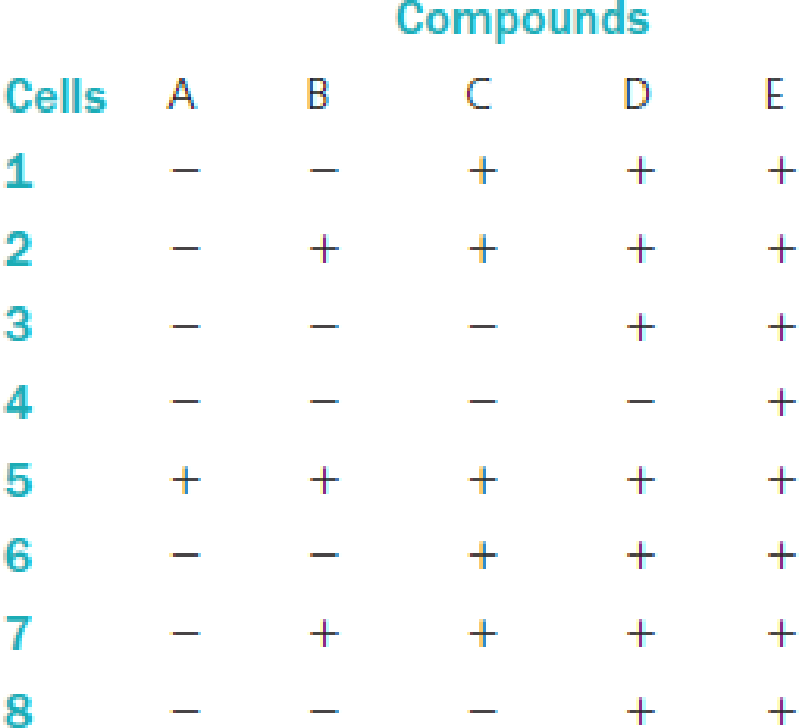
Draw the pathway leading to the production of protein E.
Expert Solution & Answer

Trending nowThis is a popular solution!

Students have asked these similar questions
Compounds A, B, C, and D are known to be intermediates in the pathway for production of protein E. To determine where the block in protein-E production occurred in each individual, the various intermediates were given to each individual’s cell in culture. After a few weeks of growth with the intermediate, the cells were assayed for the production of protein E. The results for each individual’s cells are given in the following table. A plus sign means that the protein E was produced after the cells were given the intermediate listed at the top of the column. A minus sign means that the cells still could not produce protein E even after being exposed to the intermediate at the top of the column.
a) If an individual who is homozygous for the mutation found in individual 2 and heterozygous for the mutation found in individual 4 mates with an individual who is homozygous for the mutation found in individual 4 and heterozygous for the mutation found in individual 2, what could the…
This is an SDS-PAGE gel of the protein insulin. The first lane is the molecular weight standard marker. The second lane NR is the native, non-reduced protein (MW 5.7kDa). The third lane is the protein treated with beta-mercaptoethanol. Please explain what is shown in the NR lane versus the R lane.
Various concentrations of recombinant human insulin were prepared for use standards for
an HPLC method. To verify the prepared concentrations, the samples were analyzed by
measuring the absorbance at 280 nm in a short path length (5 mm) cuvette. The molar
absorption coefficient for human insulin is approximately 5.875 x 10³ M-¹cm-¹.
a. Calculate the extinction coefficient in mL mg-¹cm-¹.
b. Calculate the concentrations of the following human insulin standards if the measured
absorbances and dilutions used are:
Standard 1
Standard 2
Abs. (at 280 nm) of Diluted Sample
0.305
0.685
Dilution
145.0 µL sample, 25.0 μL buffer
130.0 µL sample, 40.0 μL buffer
Chapter 10 Solutions
HUMAN HEREDITY (LL)-W/MINDTAP ACCESS
Ch. 10.4 - Prob. 1GRCh. 10.4 - Prob. 2GRCh. 10.7 - Prob. 1EGCh. 10.7 - Prob. 2EGCh. 10 - A couple was referred for genetic counseling...Ch. 10 - A couple was referred for genetic counseling...Ch. 10 - A couple was referred for genetic counseling...Ch. 10 - Many individuals with metabolic diseases are...Ch. 10 - Prob. 2QPCh. 10 - Enzymes have all the following characteristics...
Ch. 10 - Questions 4 through 6 refer to the following...Ch. 10 - Questions 4 through 6 refer to the following...Ch. 10 - Prob. 6QPCh. 10 - Prob. 7QPCh. 10 - Prob. 8QPCh. 10 - a. Compounds A, B, C, and D are known to be...Ch. 10 - b. Compounds A, B, C, and D are known to be...Ch. 10 - a. If an individual who is homozygous for the...Ch. 10 - Prob. 12QPCh. 10 - Suppose that in the formation of phenylalanine...Ch. 10 - If phenylalanine was not an essential amino acid,...Ch. 10 - Phenylketonuria and alkaptonuria are both...Ch. 10 - The normal enzyme required for converting sugars...Ch. 10 - Knowing that individuals who are homozygous for...Ch. 10 - Prob. 18QPCh. 10 - A person was found to have very low levels of...Ch. 10 - If an extra nucleotide is inserted in the first...Ch. 10 - Transcriptional regulators are proteins that bind...Ch. 10 - Prob. 22QPCh. 10 - Prob. 23QP
Knowledge Booster


Learn more about
Need a deep-dive on the concept behind this application? Look no further. Learn more about this topic, biology and related others by exploring similar questions and additional content below.Similar questions
- a. Why proteins are extracted to a buffered solution? Briefly describe the components of ageneralized protein extraction buffer?.b. Describe the basis of affinity chromatography in protein purification.c. What is the most appropriate method of protein elution in affinity chromatography?d. List three examples of commonly employed combinations of ligand and protein in affinitychromatography.arrow_forwardSuppose that you are tasked to determine the protein concentration of an unknown protein solution via Bradford assay. You prepared six solutions of bovine serum albumin (BSA) with different concentrations. The initial concentration of the BSA stock solution is 7.50 mg/mL. Approximately 200 µL of Bradford Reagent was added to each of these solutions and the absorbance at 595 nm was taken after 5 minutes. See the table below for data on the standard solutions. Standard # A595 BSA conc (mg/mL) 0.000 0.158 2 1.125 0.291 2.250 0.372 4 3.375 0.503 5 4.500 6 5.625 0.675 Determine the protein concentration, in mg/mL, of the unknown solution if its absorbance at 595 nm was 0.248. Note: Final answer format must be x.xx (two decimal places). Round off only in the final answer. Do not round off in the middle of calculation.arrow_forward. You need to prepare 500 μL of a 250 µg/mL bovine gamma globulin solution, for one of your protein standards in the Bradford assay. The available bovine gamma globulin in the lab is in the form of a 2 mg/mL stock solution, and it can be diluted with deionized water. Describe or show below how you would create your desired 250 µg/mL solution.arrow_forward
- A. Lineweaver-Burk plot of the enzyme with increasing amounts of substrate in the absence or the presence of the inhibitor is shown below. Graph A : x-intercept Graph B : x-intercept = - 0.012, y-intercept = 0.8 Graph C : x-intercept = - 0.027, y-intercept = 0.8 Graph D : x-intercept = - 0.039, y-intercept = 0.8 - 0.007, y-intercept = 0.8 Graph A 4 Graph B Graph C Graph D 1 -0,04 -0,02 0,00 0,02 0,04 1/[Substrate] (uM) (i) Which graph indicates an enzymatic reaction without inhibitor? (ii) Which type of inhibitor is it? Briefly explain. (iii) Which graph indicates the highest concentration of inhibitor? (iv) Calculate the Vmax and Km of the graph showing an enzymatic reaction with the lowest concentration of inhibitor. Show the steps of calculation and unit in your answers. Keep 2 decimal places in your answers. 1/Rate (umol/min)arrow_forward(b) Both laboratories used 10 micrograms of protein each in their kinetic assays. Protein concentrations weredetermined by the Bradford protein assay. Assay conditions employed in the two labs (pH, temperature,etc.) were also identical. What would be the most plausible cause for the discrepancy in the Vmax valuesfor the compound I? Explain.Recall that the Bradford assay measures total protein amounts in sample solution based on complexformation between a dye and proteins. Also, the assay solution used in both labs does not contain anyinhibitors.arrow_forwardProteinase K should be used as the first step of DNA purification (before applying phenol CIA mixture) to remove most proteins. Proteinase K alone cannot remove all proteins from the lysate. Explain why the Proteinase K enzyme cannot remove ALL proteins from the cell lysate.arrow_forward
- *The enzyme glucose oxidase isolated from the mold Penicillium notatum catalyzes the oxidation of 3-D-glucose to D-glucono-6- lactose. This enzyme is highly specific for the ß anomer of In spite of this glucose and does not affect the a anomer. specificity, the reaction catalyzed by glucose oxidase is commonly used in a clinical assay for total blood glucose that is, for solutions consisting of a mixture of 3- and a-D- glucose. What are the circumstances required to make this possible? Aside from allowing the detection of smaller quan- tities of glucose, what advantage does glucose oxidase offer over non-enzymatic oxidizing agents like Tollens reagent? *Is B-D-glucosamine a reducing sugar?arrow_forwardPyridoxal phosphate (PLP) is a coenzyme for the enzyme ornithine aminotransferase. The enzyme was purified from cells grow in PLP = deficient media as well as from cells grown in media that contained pyridoxal phosphate. The stability of the two different enzyme preparations was then measured by incubating the enzyme at 37°C for different lengths of time and then assaying for the amount of enzyme activity remaining. The following results were obtained. (a) Why does the amount of active enzyme decrease with the time of incubation? (b) Why does the amount of enzyme from the PLP deficient cells decline more rapidly?arrow_forwardtopic: Bradford AssayThere are numerous methods of protein determination in use, but this module focuses on the Bradford assay.The Bradford assay is a dye-binding method that employs Coomassie Brilliant Blue G-250, whose structureis shown in Figure 2.3.4.1. Coomassie Brilliant Blue G-250 is a dye that interacts with proteins throughhydrophobic and electrostatic interactions. What are the identities and functions of the components of the Bradford reagent in protein contentdetermination?arrow_forward
- Please provide a separation solution of combining two types of chromatography to isolate the protein A from the mixture containing protein A, B, C and D (shown as below). And then explain the related mechanisms in details. Protein A: Met-Leu-Leu-Leu-Leu-Val-Val-Val-lle-lle-lle-Leu-Leu-Leu-Leu- Protein B: Met-Asp-Glu-Glu-Glu-Glu-Asp-Asp-Asp-Glu-Glu-Asp-Asp-Glu Protein C: Met-Leu-Leu-Leu-Leu-Val-Val-Val-lle-lle-lle-Leu-Leu-Leu-Leu-Val-Val-Val- Ile-lle-lle-Leu-Leu- Leu-Leu-Val-Val-Val-lle-lle-lle-Leu-Leu-Leu-Leu-Val-Val-Val-lle-lle- lle-Leu-Leu-Leu-Leu-Val-Val-Val-Ile- Protein D: Met-Asp-Glu--Glu-Glu-Glu-Asp-Asp-Asp-Glu-Glu-Asp-Asp-Glu-Glu-Asp- Glu-Asp-Glu-Glu-Asp-Asp-Glu-Glu-Glu-Glu-Asp-Asp-Asp-Glu-Glu-Asp-Asp-Glu-Glu- Asp-Glu-Asp-Glu-Glu-Asp-Glu-Glu-Asparrow_forwardDuring SDS-PAGE, glycoproteins migrate as relatively diffuse bands, whereas nonglycosylated proteins typically migrate as narrow, well-defined bands. Explain the reason for this difference in electrophoretic behavior.arrow_forwardYou have expressed a protein of interest in E. coli cells for further study in the lab. The protein has a net positive charge at pH 6, absorbs UV light at 280m, and has insulin binding activity.Briefly describe a purification scheme with at least three steps that will leverage these properties and generate pure protein.arrow_forward
arrow_back_ios
SEE MORE QUESTIONS
arrow_forward_ios
Recommended textbooks for you
 Human Heredity: Principles and Issues (MindTap Co...BiologyISBN:9781305251052Author:Michael CummingsPublisher:Cengage Learning
Human Heredity: Principles and Issues (MindTap Co...
Biology
ISBN:9781305251052
Author:Michael Cummings
Publisher:Cengage Learning
cell culture and growth media for Microbiology; Author: Scientist Cindy;https://www.youtube.com/watch?v=EjnQ3peWRek;License: Standard YouTube License, CC-BY