
EBK ANATOMY & PHYSIOLOGY
5th Edition
ISBN: 9780321888013
Author: Hoehn
Publisher: VST
expand_more
expand_more
format_list_bulleted
Concept explainers
Videos
Textbook Question
Chapter 4, Problem 16CYU
You are looking at muscle tissue through the microscope and you see striped branching cells that connect with one another. What type of muscle are you viewing?
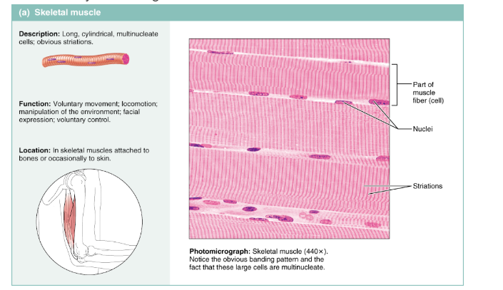
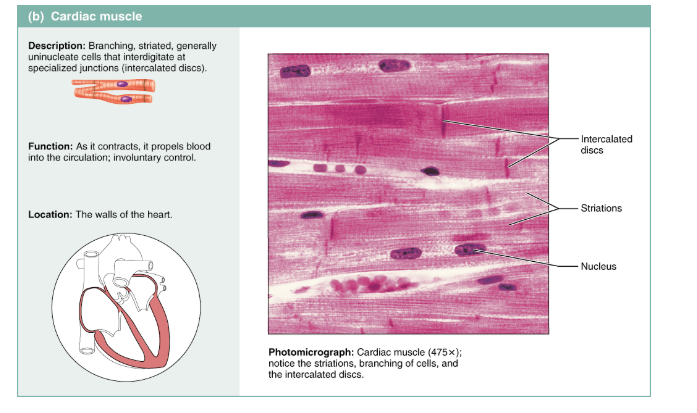
Figure 4.9 Muscle tissues.
(a) Skeletal muscle tissue. (For a related image, see A Brief Atlas of the Human Body, Plate 28.)
(b) Cardiac muscle tissue. (For a related image, see A Brief Atlas of the Human Body, Plate 31.)
View histology slides MasteringA&P® >Study Area>
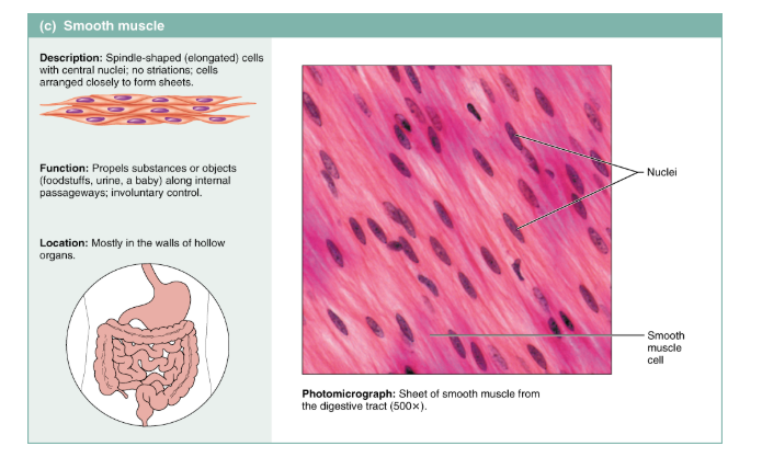
Figure 4.9 (continued) Muscle tissues.
(c) Smooth muscle tissue. (For a related image, see A Brief Atlas of the Human Body, Plate 32.)
Expert Solution & Answer
Want to see the full answer?
Check out a sample textbook solution
Students have asked these similar questions
Draw and label to show the structure of
1. Striated muscle
2. Smooth muscle
3. Heart muscle
Identify the tissues (A-D). Each letter will only be used once. Then, you will be asked for one specific characteristic that helped you to ID the tissue. Ex: spindle shape
Skeletal Muscle
How did you know this? ___________________
Cardiac Muscle
How did you know this? ___________________
Smooth Muscle
How did you know this? ___________________
Nervous Tissue
How did you know this? ___________________
Cardiac muscle differs from smooth and skeletal muscle because....
It is composed of long and cylindrical fibers, each of which has multiple nuclei.
It is composed of a branching network of fibers and is only found in vertebrates.
It typically contracts more rapidly than skeletal or smooth muscle.
It is composed of long and cylindrical fibers with one nucleus per cell.
It is composed of a branching network protected by chitin.
Chapter 4 Solutions
EBK ANATOMY & PHYSIOLOGY
Ch. 4 - Prob. 1CYUCh. 4 - Prob. 2CYUCh. 4 - Epithelial tissue is the only tissue type that has...Ch. 4 - Prob. 4CYUCh. 4 - Stratified epithelia are built for protection or...Ch. 4 - Some epithelia are pseudostratified. What does...Ch. 4 - Where is transitional epithelium found and what is...Ch. 4 - What common secretion do all unicellular exocrine...Ch. 4 - Prob. 9CYUCh. 4 - Prob. 10CYU
Ch. 4 - What are four functions of connective tissue?Ch. 4 - What are the three types of fibers found in...Ch. 4 - Which connective tissue has a soft weblike matrix...Ch. 4 - What type of connective tissue is damaged when you...Ch. 4 - Prob. 15CYUCh. 4 - You are looking at muscle tissue through the...Ch. 4 - Which muscle type(s) is voluntary? Which is...Ch. 4 - How does the extended length of a neurons...Ch. 4 - What type of membrane consists of epithelium and...Ch. 4 - What type of membrane lines the thoracic walls and...Ch. 4 - Prob. 21CYUCh. 4 - Why does a deep injury to the skin result in...Ch. 4 - Use the key to classify each of the following...Ch. 4 - An epithelium that has several layers, with an...Ch. 4 - Match the epithelial types named in column B with...Ch. 4 - The gland type that secretes products such as...Ch. 4 - The membrane which lines body cavities that open...Ch. 4 - Scar tissue is a variety of (a) epithelium, (b)...Ch. 4 - Define tissue.Ch. 4 - Name four important functions of epithelial tissue...Ch. 4 - Describe the criteria used to classify covering...Ch. 4 - Prob. 10RQCh. 4 - Provide examples from the body that illustrate...Ch. 4 - Name the primary cell type in connective tissue...Ch. 4 - Name the two major components of matrix and, if...Ch. 4 - Matrix is extracellular. How does the matrix get...Ch. 4 - Name the specific connective tissue type found in...Ch. 4 - What is the function of macrophages?Ch. 4 - Differentiate between the roles of neurons and the...Ch. 4 - Compare and contrast skeletal, cardiac, and smooth...Ch. 4 - Describe the process of tissue repair, making sure...Ch. 4 - In what ways are adipose tissue and bone similar?...
Knowledge Booster


Learn more about
Need a deep-dive on the concept behind this application? Look no further. Learn more about this topic, biology and related others by exploring similar questions and additional content below.Similar questions
- You are looking at muscle tissue through the microscope and you see striped branching cells that connect with one another. What type of muscle are you viewing?arrow_forward(a) A nerve cell's long extensions enable it to conduct electrical impulses from one body part to another. (b) The sheetlike organization of epithelial cells enables them to protect underlying cells. (c) The alignment of contractile proteins within muscle cells enables them to contract, pulling closer together the structures to which they attach. Figure 3.1 Cells vary in shape and function.arrow_forwardDuring a lab practical, a student examines a tissue that is composed of densely packed protein fibers that run parallel to each other and form a cord there are no striations, but small nuclei are visible. The student identifies the tissue as skeletal muscle. Why is the students choice wrong, and what tissue is he probably observing?arrow_forward
- Skeletal muscle structure Use the dropdown menu to select the answer that best competes each statement. Membranous channel extending inward from muscle fiber membrane (Click to select) Cytoplasm of a muscle fiber (Click to select) Connective tissue located between adjacent muscles (Click to select) Layer of connective tissue that separates a muscle into small bundles called fascicles (Click to select) v Plasma membrane of a muscle fiber (Click to select) v Layer of connective tissue that surrounds a skeletal muscle (Click to select)arrow_forwardTaking note of the parts of the muscle cells and the structural differences of the cells, W hat can you observe from the skeletal, cardiac, and smooth muscles when viewed under a micrscope?arrow_forwardand draw: 1. Obtain a slide of smooth muscle tissue from the slide box. 2. View the slide on an appropriate objective. 3. Fill out the blanks next to your drawing. 4. In the circle below, draw a representative sample of key features you identified, taking care to correctly and clearly draw their true shapes and directions. Draw your structur proportionately to their size in your microscope's field of view. Total Magnification: Type of Muscle Tissue: Smooth Muscle Tissue Source: Transwerse Surion Function of Tissue: Cey features to find nd draw: ENSES AND ATTRIBUTIONS LICENSED CONTENT, ORIGINAL A&P Labs. Authored by: Ross Whitwam. Provided by: Mississippi University for Women. Located at: http://www.muw.edu. License: CC BY-SA: Attribution-ShareAlike LICENSED CONTENT, SPECIFIC ATTRIBUTION Exercise 3.4 A. Authored by: Kent Christensen, Ph.D., J. Matthew Velkey, Ph.D., Lloyd M. Stoolman, M.D., Laura Hessle Diedra Mosley-Brower. Provided by: University of Michigan Histology and Virtual…arrow_forward
- If you will be observing cross sections of a striated muscle, how would you differentiate a skeletal muscle from a cardiac muscle?arrow_forwardwhat can you observe from the smooth, cardiac, and skeletal muscles under a microscope? (taking note of the parts of the muscle cells and structural differences of the cells)arrow_forwardWhich of the following statements can CORRECTLY be made about muscle tissue? 1. The main characteristic of muscle tissue is its ability to contract, or shortens, making movement possible. II. Skeletal muscle fibers appear banded, with many cells at the periphery and under involuntary control. II. Cardiac muscle cells are cylindrical, not striated, and have a centrally located nucleus and under involuntary IV. Smooth muscle cells are tapered at each end, are not striated, and have a single control. nucleus and under involuntary control. O Statements I and IV are correct O Statements I, II, IV are correct O Statements 1, II, HI, and IV are correct O Statements II and II are correct 21 22 23 24 25 26 27 28 29 30arrow_forward
- Skeletal muscle cell(key terms: striated, contracting proteins, contractions, nucleus location) One important structure/feature of a muscle cell isarrow_forwardIn lookingthrough a microscope how could you distinguish skeletalmuscle tissue from smooth muscle?arrow_forwardIdentify the muscle tissue types and the nervous tissue in Figure 1 _____________________________________________ 2. _____________________________________________ 3. _____________________________________________ 4. _____________________________________________arrow_forward
arrow_back_ios
SEE MORE QUESTIONS
arrow_forward_ios
Recommended textbooks for you

 Human Biology (MindTap Course List)BiologyISBN:9781305112100Author:Cecie Starr, Beverly McMillanPublisher:Cengage Learning
Human Biology (MindTap Course List)
Biology
ISBN:9781305112100
Author:Cecie Starr, Beverly McMillan
Publisher:Cengage Learning
Types of Human Body Tissue; Author: MooMooMath and Science;https://www.youtube.com/watch?v=O0ZvbPak4ck;License: Standard YouTube License, CC-BY