
Concept explainers
Videos
Marfan syndrome is an autosomal dominant disorder in humans. It results from mutation of the gene on chromosome
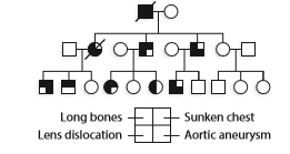
Since all cases of Marfan syndrome are caused by mutation of the fibrillin gene, and all family members with Marfan syndrome carry the same mutant allele, how do you e xplain the differences shown in the pedigree?
Want to see the full answer?
Check out a sample textbook solution
Chapter 4 Solutions
GENETIC ANALYSIS: INTEGRATED - ACCESS


- Name two ways in which loss of p53 function contributes to a malignant phenotype. Explain how benzo(a) pyrene can cause loss of p53 function. Hint: Loss of p53 function occurs in the majority of human tumors.arrow_forwardAngelica just learned that her paternal uncle, Aaron, passed away from hypertrophic cardiomyopathy (autosomal dominant). Angelica’s father was killed in a duty as a young man, and therefore, his status is unknown. Her two sisters, Eliza and Peggy, have been tested for the known causative mutation in the family and do not have it. What is the chance that Angelica has the familial mutation for HCM? Group of answer choices 1/33 1/10 1/21arrow_forwardDiscuss the following types of mutations, with reference to specific genetic disorders: i) Chromosomal deletion; ii) Reciprocal translocation; and iii) Haploinsufficiencyarrow_forward
- Loss of p53 function occurs in the majority of human tumors. Name two ways in which loss of p53 function contributes to a malignant phenotype. Explain how benzo(a) pyrene can cause loss of p53 function.arrow_forwardEhler-Danlos syndrome is a rare disorder caused by a mutation ina gene that encodes a protein called collagen (type 3 A1). Collagenis found in the extracellular matrix that plays an important role inthe formation of skin, joints, and other connective tissues. Peoplewith Ehler-Danlos syndrome have extraordinarily flexible skin and very loose joints. The pedigree below contains several individualsaffected with this syndrome, shown with black symbols. Based onthis pedigree, does the syndrome appear to follow autosomalrecessive, autosomal dominant, X-linked recessive, or X-linkeddominant inheritance? Explain your reasoning.arrow_forwardHurler syndrome is due to a mutation in a gene that encodes aprotein called α-l-iduronidase. This protein functions withinlysosomes as an enzyme that breaks down mucopolysaccharides(a type of polysaccharide that has many acidic groups attached).When this enzyme is defective, excessive amounts of the mucopolysaccharides dermatan sulfate and heparin sulfate accumulatewithin the lysosomes, especially in liver cells and connectivetissue cells. This accumulation leads to symptoms such as anenlarged liver and spleen, bone abnormalities, corneal clouding,heart problems, and severe neurological problems. The pedigreebelow contains three members affected with Hurler syndrome,indicated with black symbols. Based on this pedigree, does thissyndrome appear to follow autosomal recessive, autosomaldominant, X-linked recessive, or X-linked dominant inheritance?Explain your reasoning.arrow_forward
- Sickle cell disease— what is the background of this disorder: include the name of the disorder, any alternate names used, provide a description of the disorder, and the typical age of onset. What is the Type of genetic disorder: explain if and how this disorder is inherited. Explain the genetic causes of the disorder. Is it inherited? Is the disorder a dominant or recessive trait? Which chromosome is affected in this disorder? Is a gene mutated? If so, name the gene that is affected? How is gene expression impacted (is it a particular type of mutation, a case of a misshapen protein, etc.?)arrow_forwardTalk about the challenges involved in determining the genetic components of polygenic illnesses. Explain complementation groups and how the biochemical underpinnings of disease are determined using them. Hereditary illnesses of genomic instability include Werner syndrome, Bloom syndrome, XP, ataxia-telangiectasia, and Fanconi anemia. Which of these ailments has molecular mechanisms behind it? Which kind of genetic instability is connected to which disorder?.arrow_forwardGene mutations can be classified in two major ways:(1) hereditary or germline mutations that are inherited from a parent and are present throughout a person’s life in virtually every cell in the body.(2) acquired or somatic mutations that occur at some time during a person’s life and are present only in certain cells, not in every cell in the body.If there is no family history of a particular disease but a child has the disease then it may have arisen due to a(n) ________ mutation early during development. A) acquired B) inherited C) silent D) transitionarrow_forward
- Junctional epidermolysis bullosa (JEB) is a severe skin disorder that results in blisters over the entire body. The disorder is caused by autosomal recessive mutations at any one of three loci that help to encode laminin 5, a major component in the dermal–epidermal basement membrane. Leena Pulkkinen and colleagues described a male newborn who was born with JEB and died at 2 months of age (L. Pulkkinen et al. 1997. American Journal of Human Genetics 61:611–619); the child had healthy, unrelated parents. Chromosome analysis revealed that the infant had 46 normal-appearing chromosomes. Analysis of DNA showed that his mother was heterozygous for a JEB-causing allele at the LAMB3 locus, which is on chromosome 1. The father had two normal alleles at this locus. DNA fingerprinting demonstrated that the male assumed to be the father had, in fact, conceived the child. Q. Assuming that no new mutations occurred in this family, explain the presence of an autosomal recessive disease in the child…arrow_forwardJunctional epidermolysis bullosa (JEB) is a severe skin disorder that results in blisters over the entire body. The disorder is caused by autosomal recessive mutations at any one of three loci that help to encode laminin 5, a major component in the dermal–epidermal basement membrane. Leena Pulkkinen and colleagues described a male newborn who was born with JEB and died at 2 months of age (L. Pulkkinen et al. 1997. American Journal of Human Genetics 61:611–619); the child had healthy, unrelated parents. Chromosome analysis revealed that the infant had 46 normal-appearing chromosomes. Analysis of DNA showed that his mother was heterozygous for a JEB-causing allele at the LAMB3 locus, which is on chromosome 1. The father had two normal alleles at this locus. DNA fingerprinting demonstrated that the male assumed to be the father had, in fact, conceived the child. Q. How might you go about proving your explanation? Assume that a number of genetic markers are available for each chromosome.arrow_forwardWhat is the Philadelphia chromosome? Briefly describe how it causes chronic myeloid leukemia.arrow_forward
 Human Anatomy & Physiology (11th Edition)BiologyISBN:9780134580999Author:Elaine N. Marieb, Katja N. HoehnPublisher:PEARSON
 Biology 2eBiologyISBN:9781947172517Author:Matthew Douglas, Jung Choi, Mary Ann ClarkPublisher:OpenStax
 Anatomy & PhysiologyBiologyISBN:9781259398629Author:McKinley, Michael P., O'loughlin, Valerie Dean, Bidle, Theresa StouterPublisher:Mcgraw Hill Education,
 Molecular Biology of the Cell (Sixth Edition)BiologyISBN:9780815344322Author:Bruce Alberts, Alexander D. Johnson, Julian Lewis, David Morgan, Martin Raff, Keith Roberts, Peter WalterPublisher:W. W. Norton & Company
 Laboratory Manual For Human Anatomy & PhysiologyBiologyISBN:9781260159363Author:Martin, Terry R., Prentice-craver, CynthiaPublisher:McGraw-Hill Publishing Co.
 Inquiry Into Life (16th Edition)BiologyISBN:9781260231700Author:Sylvia S. Mader, Michael WindelspechtPublisher:McGraw Hill Education