
Laboratory Manual for Human Anatomy & Physiology Main Version
4th Edition
ISBN: 9781260159110
Author: Terry Martin
Publisher: Mcgraw-hill Higher Education (us)
expand_more
expand_more
format_list_bulleted
Videos
Textbook Question
Chapter 5, Problem 1.2A
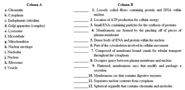
Match the cellular components in column A with the descriptions in column B- Place the letter of your choice in the space provided.
Expert Solution & Answer
Want to see the full answer?
Check out a sample textbook solution
Students have asked these similar questions
Identify the cell indicated by the arrow
Identify the cell
Write a sentence or two defining the essential function or structure where
appropriate of the following cell parts.
Chapter 5 Solutions
Laboratory Manual for Human Anatomy & Physiology Main Version
Ch. 5 - Which of the following cellular structures is not...Ch. 5 - Which of the following cellular structures is...Ch. 5 - The outer boundary of a cell is the mitochondrial...Ch. 5 - Microtubules, intermediate filaments, and...Ch. 5 - Easily attainable living cells observed in this...Ch. 5 - A slide of human cheek cells can be stained to...Ch. 5 - Cellular energy is stored in ER. ATP. DNA. RNA.Ch. 5 - The smooth ER possesses ribosomes. True...Ch. 5 - The nuclear envelope contains nuclear pores. True...Ch. 5 - The cells lining the inside of the cheek are...
Ch. 5 - Figure 5.4 Label the indicated cellular structure...Ch. 5 - Match the cellular components in column A with the...Ch. 5 - Prob. 2.1ACh. 5 - Prob. 2.2ACh. 5 - What do the various types of cells in these...Ch. 5 - What are the main differences you observed among...Ch. 5 - Prob. 3.4ACh. 5 - Electron micrographs represent extremely thin...Ch. 5 - Electron micrographs represent extremely thin...Ch. 5 - Electron micrographs represent extremely thin...Ch. 5 - Electron micrographs represent extremely thin...Ch. 5 - Electron micrographs represent extremely thin...Ch. 5 - Electron micrographs represent extremely thin...Ch. 5 - Electron micrographs represent extremely thin...Ch. 5 - Electron micrographs represent extremely thin...Ch. 5 - Electron micrographs represent extremely thin...Ch. 5 - Electron micrographs represent extremely thin...
Additional Science Textbook Solutions
Find more solutions based on key concepts
Why is it unlikely that two neighboring water molecules would be arranged like this?
Campbell Biology (11th Edition)
Which of the following would be used to identify an unknown bacterial culture that came from a patient in the i...
Microbiology Fundamentals: A Clinical Approach
The correct term for production of offspring. Introduction: Reproduction is an important life process for most ...
Biology Illinois Edition (Glencoe Science)
What are the cervical and lumbar enlargements?
Principles of Anatomy and Physiology
Define histology.
Fundamentals of Anatomy & Physiology (11th Edition)
Knowledge Booster


Learn more about
Need a deep-dive on the concept behind this application? Look no further. Learn more about this topic, biology and related others by exploring similar questions and additional content below.Similar questions
- _______ _________ Are plant and animal cells with a nucleus and membrane enclosed organelles. fill in the blankarrow_forwardWrite a short description for each of the cells listed below that would help a student recognize that cell type from a picture or a microscope view. For each description, use five words or less. (Note that you will need to think about the most distinguishing and important features of the cells to describe them in so few words.) 1. squamous epithelial cells 2. red blood cells 3. white blood cells 4. skeletal muscle cells 5. cartilage cells 6. nerve cellsarrow_forwardb. Cell Type: How many layers do you see?arrow_forward
arrow_back_ios
SEE MORE QUESTIONS
arrow_forward_ios
Recommended textbooks for you

Animal Communication | Ecology & Environment | Biology | FuseSchool; Author: FuseSchool - Global Education;https://www.youtube.com/watch?v=LsMbn3b1Bis;License: Standard Youtube License