
Laboratory Manual for Human Anatomy & Physiology Main Version
4th Edition
ISBN: 9781260159110
Author: Terry Martin
Publisher: Mcgraw-hill Higher Education (us)
expand_more
expand_more
format_list_bulleted
Concept explainers
Videos
Textbook Question
Chapter 5, Problem F5.4A
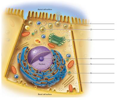
Figure 5.4 Label the indicated cellular structure of this composite cell.
Expert Solution & Answer
Want to see the full answer?
Check out a sample textbook solution
Students have asked these similar questions
Identify the shape of the cell indicated by the arrow
Identify the light blue structure in the center of all cell
Label the cell diagram
Chapter 5 Solutions
Laboratory Manual for Human Anatomy & Physiology Main Version
Ch. 5 - Which of the following cellular structures is not...Ch. 5 - Which of the following cellular structures is...Ch. 5 - The outer boundary of a cell is the mitochondrial...Ch. 5 - Microtubules, intermediate filaments, and...Ch. 5 - Easily attainable living cells observed in this...Ch. 5 - A slide of human cheek cells can be stained to...Ch. 5 - Cellular energy is stored in ER. ATP. DNA. RNA.Ch. 5 - The smooth ER possesses ribosomes. True...Ch. 5 - The nuclear envelope contains nuclear pores. True...Ch. 5 - The cells lining the inside of the cheek are...
Ch. 5 - Figure 5.4 Label the indicated cellular structure...Ch. 5 - Match the cellular components in column A with the...Ch. 5 - Prob. 2.1ACh. 5 - Prob. 2.2ACh. 5 - What do the various types of cells in these...Ch. 5 - What are the main differences you observed among...Ch. 5 - Prob. 3.4ACh. 5 - Electron micrographs represent extremely thin...Ch. 5 - Electron micrographs represent extremely thin...Ch. 5 - Electron micrographs represent extremely thin...Ch. 5 - Electron micrographs represent extremely thin...Ch. 5 - Electron micrographs represent extremely thin...Ch. 5 - Electron micrographs represent extremely thin...Ch. 5 - Electron micrographs represent extremely thin...Ch. 5 - Electron micrographs represent extremely thin...Ch. 5 - Electron micrographs represent extremely thin...Ch. 5 - Electron micrographs represent extremely thin...
Knowledge Booster


Learn more about
Need a deep-dive on the concept behind this application? Look no further. Learn more about this topic, biology and related others by exploring similar questions and additional content below.Similar questions
- 1 2- 3 4 5 6 Label the indicated structures in each type of cell in the illustrations below. Centrosome 15 14 13 Wall of adjacent cell AZA Plasmodesmata 12 8 -9 -10 11 12 13 -14 15arrow_forwardin only one cell, label the nucleus, cytoplasm, and cell membrane for these two figures. Describethese cells based on their cell shape.arrow_forwardShape of cellarrow_forward
- Give the actual dimensions (lenght and width) of the cell shown in this microscope drawing. You are told that. the depth of this type of squamous cell is typically 460nm. Use this information to calculate the volume of the cell.arrow_forwardLabel the organelles in this cell.arrow_forwardUsing the letters from column B, match the cell description in column A. (Note that all require more than a single choice.)arrow_forward
- Match the following cell structures with their descriptions. 1. Fibers of the cytoskeleton that attach to chromosomes and move them during mitosis 2. Cell junctions that seal cells so tightly together that materials cannot pass between the cells Cilia Intermediate filaments 3. Cell surface appendages that contain microtubules and beat to move substances across the surface Tight junctions of a cell 4. The network of many types of protein fibers that gives shape to the cell and anchors the organelles Microtubules Desmosomes 5. Cell junctions that link the cytoskeleton of adjacent cells in order to prevent the cells from being pulled apart Microfilaments/actin filaments 6. Fibers of the cytoskeleton that allow cells such as amoebae to crawl aroundarrow_forwardDrag and drop to identify the organelles of this cell.arrow_forwardFind and label the cell wall, cytoplasm, vacuole bounded by tonoplast, nucleus (if visible) of this turgid cell in the picture below. pls answer tysm!arrow_forward
arrow_back_ios
SEE MORE QUESTIONS
arrow_forward_ios
Recommended textbooks for you

Biology - Intro to Cell Structure - Quick Review!; Author: The Organic Chemistry Tutor;https://www.youtube.com/watch?v=vwAJ8ByQH2U;License: Standard youtube license