Hole's Human Anatomy & Physiology
15th Edition
ISBN: 9781259864568
Author: SHIER, David, Butler, Jackie, Lewis, Ricki
Publisher: Mcgraw-hill Education,
expand_more
expand_more
format_list_bulleted
Concept explainers
Videos
Textbook Question
Chapter 5, Problem 4IA
Answer the following questions with respect to the micrographbelow (80). 
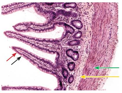
(a) Identify the organ depicted.
(b) What type oftissue is depicted (green arrow, yellow arrow)? (c) To what celldoes the arrow point (red arrow, black arrow)?

Expert Solution & Answer
Want to see the full answer?
Check out a sample textbook solution
Students have asked these similar questions
Identify A (arrows; name of the area; blank 1) - Identify B (arrow; name of
the structure, blank 2) - Identify C (arrows; name of the cells, blank 3) - D: Identify
the organ (blank 4)
Low
high. mag
D: Identify this organ
Blank # 1
Blank # 2
Blank # 3
Blank # 4
Identify A (name of the layer; blank 1) - Identify B (name of the structure; blank 2) -
Identify C (name of the layer; blank 3) - Identify D (name of the cells; blank 4) - E:
identify this organ.
High mag.
Low
high mag.
D.
B
B
E: Identify this organ
Blank # 1
Blank # 2
Blank # 3
Blank # 4
Blank # 5
Identify these cells (white arrow)
Choose one from the following:
(A) hepatocytes
(B) enteroendocrine cells
(C) Paneth cells
(D) acinar cells
(E) alpha/beta cells
(F) Kupffer cells
Chapter 5 Solutions
Hole's Human Anatomy & Physiology
Ch. 5 - Prob. 1PCh. 5 - Prob. 2PCh. 5 - Prob. 3PCh. 5 - Prob. 4PCh. 5 - Prob. 5PCh. 5 - Prob. 6PCh. 5 - Prob. 7PCh. 5 - Prob. 8PCh. 5 - Prob. 9PCh. 5 - Prob. 10P
Ch. 5 - What are the general characteristics of connective...Ch. 5 - Prob. 12PCh. 5 - Prob. 13PCh. 5 - Prob. 14PCh. 5 - Prob. 15PCh. 5 - Prob. 16PCh. 5 - Prob. 17PCh. 5 - Prob. 18PCh. 5 - Prob. 19PCh. 5 - Prob. 20PCh. 5 - Name the four types of membranes, and explain how...Ch. 5 - Prob. 22PCh. 5 - Distinguish among skeletal, smooth, and cardiac...Ch. 5 - Prob. 24PCh. 5 - Prob. 25PCh. 5 - 1 Define tissue. (p. 149)
Ch. 5 - Prob. 2CACh. 5 - Prob. 3CACh. 5 - Prob. 4CACh. 5 - Prob. 5CACh. 5 - Prob. 6CACh. 5 - Prob. 7CACh. 5 - Prob. 8CACh. 5 - Prob. 9CACh. 5 - Prob. 10CACh. 5 - Prob. 11CACh. 5 - Prob. 12CACh. 5 - Prob. 13CACh. 5 - Prob. 14CACh. 5 - Prob. 15CACh. 5 - Prob. 16CACh. 5 - Contrast dense regular and dense irregular...Ch. 5 - Prob. 18CACh. 5 - Prob. 19CACh. 5 - Prob. 20CACh. 5 - Prob. 21CACh. 5 - Prob. 22CACh. 5 - Prob. 23CACh. 5 - Prob. 24CACh. 5 - Prob. 25CACh. 5 - Prob. 26CACh. 5 - Compare and contrast skeletal, smooth, and cardiac...Ch. 5 - Prob. 28CACh. 5 - Prob. 29CACh. 5 - Prob. 1IACh. 5 - Prob. 2IACh. 5 - Prob. 3IACh. 5 - Answer the following questions with respect to the...Ch. 5 - Prob. 5IACh. 5 - Prob. 6IACh. 5 - Prob. 7IACh. 5 - Prob. 8IA
Knowledge Booster


Learn more about
Need a deep-dive on the concept behind this application? Look no further. Learn more about this topic, biology and related others by exploring similar questions and additional content below.Similar questions
- Define the following terms: (a) Aestivation (b) Placentation (c) Actinomorphicarrow_forwardThe three micrographs on the right (A, B, C) are higher magnification of areas (labeled A, B, C, respectively) of the section on the micrograph on the left. Identify A: name of the layer/area (blank 1) - Identify B: name of the cells (blank 2) -Identify C: name of the layer/area (blank 3) - Identify D: name of the organ. Low high. mag A D: Identify this organarrow_forwardIdentify the four kinds of tissue membranes shown in the drawing below.arrow_forward
- What is the tissue labeled A?arrow_forwardComplete the following: After comparing the wet mount and the stained cheek cells, state the advantage that was gained by staining cells...............................arrow_forward(a) From which blood cells do microglial cells originate? (b) What are the functions of microglial cells?arrow_forward
- 10:41 task connective ti... 1. Identify the fabric on the prepared preparation. Name the Stain. Describe the histological structures of the preparation, indicated by numbers (1-2.3). List the main functions 2. identify connective tissue cells 3. Identify the type on the prepared preparation. Name the Stain. Describe the histological structures of the preparation, indicated by numbers (1-2.3). List the main functions ...arrow_forwardDefine pleitropyarrow_forwardDefine frenulumarrow_forward
- What is tissue harmonic imaging?arrow_forwardMatch the following questionsarrow_forwardFor each of the lettered regions identified on this figure, giveits name and function.(a) __________________________(b) __________________________(c) __________________________(d) __________________________(e) __________________________(f) __________________________(g) __________________________(h) __________________________(i) __________________________(j) __________________________arrow_forward
arrow_back_ios
SEE MORE QUESTIONS
arrow_forward_ios
Recommended textbooks for you

Types of Human Body Tissue; Author: MooMooMath and Science;https://www.youtube.com/watch?v=O0ZvbPak4ck;License: Standard YouTube License, CC-BY