
Concept explainers
Videos
Answers to all problems are at the end of this book. Detailed solutions are available in the Student Solutions Manual, Study Guide, and Problems Book.
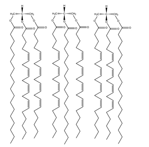
Drawing Structures of Triacylglycerols Draw the structures of (a) all the possible triacylglycerols that can be formed from glycerol with Stearic and arachidonic acid and (b) all the phosphatidylserine isomers that tan be formed from palmitic and linolenic acids.
(a)
To explain:
The formation from glycerol from stearic and arachidonic acid should be described.
Introduction:
Glycerol is the colorless, viscous liquids that belong to the organic group of alcohol. It has three carbon atoms and three hydroxyl groups in its structure that are known as trihydroxyl sugar alcohol.
Explanation of Solution
The chiral molecule is found in the second carbon of glycerol if there is the presence of different substitutes at both ends of carbon. This can lead to the formation of six triacylglycerols from stearic and arachidonic acids as below:
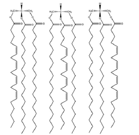
(b)
To explain:
The formation from phosphatidylserine isomers from palmitic and linolenic acids should be described.
Introduction:
A phospholipid with fatty acid is known as phosphatidylserine that covers and protects the cells in the brain and carries messages. It has two fatty acids attached with ester linkage to the C-1 and C-2 of glycerol and serine attached by a phosphodiester linkage to the C-3 of glycerol.
Explanation of Solution
There is a presence of phosphate bonded with a serine residue at C-3 of glycerol in structures of phosphatidylserine. There are two common forms of linolenic acid which means four different phosphatidylserine can be formed from palmitic and linolenic acids as shown below:

Want to see more full solutions like this?
Chapter 8 Solutions
Owlv2, 4 Terms (24 Months) Printed Access Card For Garrett/grisham's Biochemistry Technology Update, 6th


- Answers to all problems are at the end of this book. Detailed solutions are available in the Student Solutions Manual, Study Guide, and Problems Book. Determining the Branch Points and Reducing Ends of Amylopectin A 0.2-g sample of amylopectin was analyzed to determine the fraction of the total glucose residues, that are branch points in the structure. The sample was exhaustively methylated and then digested, yielding 50-mol of 2,3-dimethylgluetose and 0.4 mol of 1,2,3,6- letramethylglucose. What fraction of the total residues are branch points? I low many reducing ends does this sample of amylopectin have?arrow_forwardAnswers to all problems are at the end of this book. Detailed solutions are available in the Student Solutions Manual, Study Guide, and Problems Book. Draw the Titration Curve for a Weak Acid and Determine its pKa from the Titration Curve When a 0.1 M solution of a weak acid was titrated with base, the following results were obtained: Plot the results of this titration and determine the pK a of the weak acid from your graph.arrow_forwardAnswers to all problems are at the end of this book Detailed solutions are available in the Student Solutions Manual, Study Guide, and Problems Book. Solving the Sequence of an Oligopeptide From Sequence Analysis Data Analysis of the blood of a catatonic football fan revealed large concentrations of a. psychologic octapeptide. Amino acid analysis of this oclapeplide gave the following results: 2 Ala lArg 1 Asp 1 Mel 2 Tyr I Val 1NH/ The following facts were observed: Partial acid hydrolysis of the octapeptide yielded a dipeptide of the structure Chymolrypsin treatment of the octapeplide yielded two tetrapeptides, each containing an alanine residue. Trypsin treatment of one of the tetrapeptides yielded two dipeptides. Cyanogen bromide treatment of another sample of the same tetrapeplide yielded a tripeplideand free Tyr. N-lerminal analysis of the other tetrapeptide gave Asn. What is the amino acid sequence of this oclapeplide?arrow_forward
- Answers to all problems are at the end of this book. Detailed solutions are available in the Student Solutions Manual, Study Guide, and Problems Book. Plot the Titration Curve for Bicine and Calculate How to Prepare a pH 7.5 Bicine Buffer Solution Bicine (N, N—bis (2-hydroxyethyl) glycine) is another commonly used buffer in biochemistry labs. The structure of Bicine in its fully protonated form is shown here: Draw the titration curve for Bicine. assuming the pA'a for its free COOH group is 2.3 and the pAa for its tertiary amino group is 8.3. Draw the structure of the fully deprotonated form (completely dissociated form) of bicine. You have available a U.l Msolution of Bicine at its isoelectric point (pH|)T 0.1 M solutions of HCI and NaOH. and ample distilled water. Describe the preparation of 1 L of 0.U4 M Bicine buffer. pH 7.5. What is the concentration of the fully protonated form of Bicine in your final buffer solution?arrow_forwardAnswers to all problems are at the end of this book. Detailed solutions are available in the Student Solutions Manual, Study Guide, and Problems Book. Understanding Stereochemical Transformations of Amino Acids Absolute configurations of the amino acids are referenced to D- and L-glyceraldehyde on the basis of chemical transformations that can convert the molecule of interest to either of these reference isomeric structures. In such reactions, the stereochemical consequences for the asymmetric centers must be understood for each reaction step. Propose a sequence of reactions that would demonstrate that L(-)-serine is stereochemically related to l(- )-glyceraldehyde.arrow_forwardAnswers to all problems are at the end of this book.. Detailed solutions are available in the Student Solutions Manual, Study Guide, and Problems Book. Calculating the Composition of Anomeric Sugar Mixtures -D-Glucose has a specific notation, []220, of + 112.20. whereas -D-glucose has a specific notation of +18.70. What is the composition of a mixture of - and -D-glucose, which has a specific notation of 83 .U0?arrow_forward
- Answers to all problems are at the end of this book. Detailed solutions are available in the Student Solutions Manual, Study Guide, and Problems Book. Assessing the pH Dependence of Poly-L-Glutamate Structure Poly-L glutamate adopts an tr-helical structure at low pH but becomes a random coil above pH 5. Explain this behavior.arrow_forwardAnswers to all problems are at the end of this book. Detailed solutions are available in the Student Solutions Manual, Study Guide, and Problems Book. (Research Problem) The Nature and Roles of Linear Motifs in Proteins In addition to domains and modules, there are other significant sequence patterns in proteins—known as linear motifs—that are associated with a particular function. Consult the biochemical literature to answer the following questions: 1. What are linear motifs? 2. How are they different from domains?. 3. What are their functions? 4. How can they be characterized? 5. There are several papers that are good starting points for this problem. Neduva, V., and Russell, R., 2005. Linear motifs: evolutionary interaction switches. FEBS Letters 579:3342-3345. Gibson, T., 2009. Cell regulation: determined to signal discrete cooperation. Trends in Biochemical Sciences 34:471-482. Diella, K. Haslam, N., Chica., C. et aL, 2009. Understanding eukaryotic linear motifs and their role in cell signaling and regulation. Frontiers of Bioscience 13:6580-6603.arrow_forwardAnswers to all problems are at the end of this book. Detailed solutions are available in the Student Solutions Manual, Study Guide, and Problems Book. Interpreting Kinetics Experiments from Graphical Patterns The following graphical patterns obtained from kinetic experiments have several possible interpretations depending on the nature of the experiment and the variables being plotted. Give at least two possibilities for each.arrow_forward
- Answers to all problems are at the end of this book. Detailed solutions are available in the Student Solutions Manual, Study Guide, and Problems Book. A Rule of Thumb for Amino Acid Content in Proteins The simple average molecular weight of the 20 common amino adds is 138, but most biochemists use 110 when estimating the number of amino acids in a protein of known molecular weight. Why do you Suppose this is? (Hint: There are two contributing factors to the answer. One of them will be apparent from a brief consideration of the amino acid compositions of common proteins. See, for example, Figure 5.16 of this text.)arrow_forwardAnswers to all problems are at the end of this book. Detailed solutions are available in the Student Solutions Manual, Study Guide, and Problems Book. Use examples from the ActiveModel for Human GaleLtin-1 to describe the hydrophobic effect.arrow_forwardAnswers to all problems are at the end of this book. Detailed solutions are available in the Student Solutions Manual, Study Guide, and Problems Book. Understanding the Phase Transitions of Membrane Phospholipids Discuss the effects on the lipid phase transition of pure dimyristoyl phosphatidylcholine vesicles of added (a) divalent cations, (b) cholesterol, (c) distearoyl phosphatidylscrine. (d) dioleoyl phosphatidylcholine, and (e) integral meinbrane proteins.arrow_forward
 BiochemistryBiochemistryISBN:9781305577206Author:Reginald H. Garrett, Charles M. GrishamPublisher:Cengage Learning