Concept explainers
Figure 18.13 Are you able to recognize and identify the various formed elements? You will need to do this is a systematic manner, scanning along the image. The standard method is to use a grid, but this is not possible with this
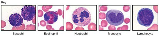
Figure 18.13 Leukocytes (Micrographs provided by the Regents of University of Michigan Medical School © 2012)

Trending nowThis is a popular solution!

Chapter 18 Solutions
Anatomy & Physiology
Additional Science Textbook Solutions
College Physics
Human Anatomy & Physiology (11th Edition)
Microbiology: An Introduction
Campbell Biology in Focus
Campbell Essential Biology with Physiology (5th Edition)
Microbiology: An Introduction (13th Edition)


- Use Figure 37.2 to identify the leukocytes in the micrograph on the left. __ neutrophil ___ lymphocyte __ eosinophil ____ basophilarrow_forwardWhy do erythrocytes on the slide appear as circular structures with pale centres?arrow_forwardin these two images identify if there are platelets and nRBcarrow_forward
- in these image identify if there are platelets and nRBCarrow_forwardA slide of blood cells is referred to as a smear or a film,why?arrow_forwardMatch the names of the blood cells in column B with their descriptions in column A. Some names are used more than once. Column A Column B(1) destroys parasites (a) erythrocyte(2) has two types of granules (b) neutrophil(3) does not use diapedesis (c) eosinophil(4) stops the allergic response (d) basophil (e) lymphocyte (f) monocyte (5) the only cell that is not spherical (6) granulocyte that phagocytizes bacteria (7) the largest blood cell (8) granulocyte with the smallest granules…arrow_forward
- Question #1: Explain ABO Blood group. Question #2: Identify and describe the cellular and non cellular components of blood Please explain in detailarrow_forwardNeutrophils and macrophages are both leukocytes - white blood cells. Describe two additional similarities and two differences between these cellsarrow_forwardWhile taking a clinical laboratory class, Marilyn prepared and examined blood smears from several donors. One of the smears had an increased percentage (about 10% of observed leukocytes) of cells containing reddish-orange granules. Discuss the type of cell described and the condition that may have caused this increase in the donor.arrow_forward
- Based on its chronology, arranged the following processes for hemostasis _______ Platelet adheres to extracellular matrix _______ Counter-regulatory mechanism _______ Platelet aggregation _______ Vascular injury _______ Release of granules, ADP and thromboxane A _______ Local activation of coagulation cascade _______ Secondary hemostasis _______ Activation of platelets _______ Transient vasoconstriction _______ Primary hemostatic plug _______ Fibrin polymerizationarrow_forwardIf the patient had a blood type different from O+, which one wouldnot have resulted in his death? (Explain your answer.)a. type A− b. type B+ c. type AB− d. type O−arrow_forwardIn hematology laboratory, automation has been the ongoing trend in terms of cell sorting and identification. Do you think it is about time to phase out the manual method of blood cell counting and identification in this area? Why or why not?arrow_forward
 Anatomy & PhysiologyBiologyISBN:9781938168130Author:Kelly A. Young, James A. Wise, Peter DeSaix, Dean H. Kruse, Brandon Poe, Eddie Johnson, Jody E. Johnson, Oksana Korol, J. Gordon Betts, Mark WomblePublisher:OpenStax College
 Comprehensive Medical Assisting: Administrative a...NursingISBN:9781305964792Author:Wilburta Q. Lindh, Carol D. Tamparo, Barbara M. Dahl, Julie Morris, Cindy CorreaPublisher:Cengage Learning