
Concept explainers
In GWAS analysis, because of the existence of LD blocks (or haplotype blocks), it is not necessary to genotype a person for every one of the 50 million known SNPs. Haplotype blocks are stretches of DNA containing particular SNP variants that tend to be inherited together (as a block) because recombination within the region is rare. In the accompanying figure, three different SNP loci are shown at top (SNP10, SNP11, and SNP12), each with two alleles among the world’s population of humans.
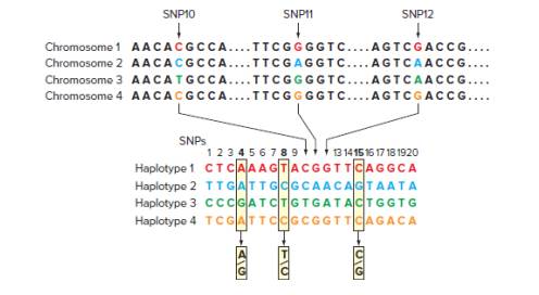
Only four of all the possible combinations of these SNP alleles are found in human genomes, as shown in the four chromosome types pictured. These three SNPs are part of a larger block of 20 SNPs that are usually inherited in one of the four configurations, or haplotypes, shown. Because these 20 SNPs are inherited as haplotype blocks, genotyping any individual for the three so-called Tag SNPs (SNP4, SNP8, and SNP15 shown in bold) should be sufficient to predict that individual’s alleles for the other 17 SNPs.
| a. | How many configurations of the three SNPs shown at the top of the diagram (SNPs 10, 11, and 12) are theoretically possible? |
| b. | How many haplotype variants are theoretically possible considering all 20 SNPs in the haplotype block, and assuming that each of them has two possible alleles? |
| c. | Given that humans are diploid, every individual has two copies of every (autosomal) haplotype block, one on each homolog. Does heterozygosity for the haplotype blocks interfere with genotyping individuals using the Tag SNPs shown in the diagram? Explain? |
| d. | In part (c), you saw that the three Tag SNPs shown in the diagram are sufficient to type any individual for this particular haplotype block. Is this the only set of three Tag SNPs that could be used? |
Want to see the full answer?
Check out a sample textbook solution
Chapter 22 Solutions
ND STONY BROOK UNIVERSITY LOOSELEAF GENETICS: FROM GENES TO GENOMES
Additional Science Textbook Solutions
Principles of Anatomy and Physiology
Human Anatomy & Physiology
Biology Illinois Edition (Glencoe Science)
Biology: Life on Earth (11th Edition)
BIOLOGY:THE ESSENTIALS (LL) W/CONNECT


- Imagine a 9-armed, hairy monster, who lives in Monster Land. It has a genome that is very similar to a mammal genome (diploid, same type of sex determination, etc). There are three different colours of this monster: Purple, Yellow, and Purple and Yellow mixed. Here is a table of 20 individuals and 10 SNPs, where the name of the SNP is the chromosome and the position (chromosome.position). Using this table, do a GWAS with only these 10 SNPs. How many markers of the genome are significantly associated with the trait? Phenotype chr1.1 chr2.8 chr3.12 chr4.12 chr4.55 chr7.88 chr9.55 chr10.1 chrX.1 chrX.5 Purple AA AT F GG CC CC 8 TT TT TT CT Purple AA AT TT GC CC CC AT TT TT CT Yellow AA AT TT GC CC CC AT AT GG CT Purple and AA TT TT GC TT CC AT AT GT CT Yellow Purple AA AT AA GG Purple AA AT 순 E TT GC AT TT TT CT AA GG TT GC AT TT TT CT Purple AA AT AA CC TT GG AT TT TT TT Purple AA AT AA GG CC GG AT TT TT TT Purple and AA TT AT CC CC GG AT AT GT TT Yellow Purple AA AT AT CC CC GC AT TT TT…arrow_forwardThe DNA of every individual in the pedigree shown in image B (below) has been sequenced at the causative locus, all the non- shaded individuals are wild type apart from III.1 and III.6. III.1 and III.6 have both been proven to have the causative allele for the condition but they do not exhibit any of the phenotypic signs or symptoms. Based on this pedigree, what is the level of penetrance for the condition? Please give your answer as a percentage to one decimal place, give the number only, no percentage symbol. ANSWER: Given the information above I calculate the level of penetrance seen in image B to be Blank 1 percent. A KEY Homozygous Homozygous Heterozygous Heterozygous Wild Type Male Female Male Female Male Note: Completely red symbol denotes an individual exhibiting the phenotype of interest CI || III IV V 1/4 1/2 1/2 1/2 1/2 Wild Type Female 1/4 1/2 Affected Known carrier Affected female Normal female Affected male Normal male D ●●●arrow_forwardBelow is a pedigree showing transmission of a disease; affected individuals are indicated by solid circles or boxes. Represented below the pedigree are two nylon membranes; DNA samples from each individual in the pedigree were spotted in identical fashion on each membrane. Each individual's DNA appears directly below his or her position in the pedigree. Each membrane was hybridized with allele specific oligonucleotides that detect the two alternative alleles of a single SNP locus (ASO 1 and ASO 2). Positive hybridization is represented as a filled circle and lack of hybridization is represented as an open circle. I 11 III IV V ASO 1 ASO 2 To 1 2 3 4 5 6 a. What is the mode of transmission of this disease? 12 b. Which SNP allele (ASO1 or ASO2) is originally linked to the disease gene? c. Draw a diagram of the event that gave rise to the genotype of individual IV-5. d. Individual V-1 is an unborn fetus. If the SNP locus is 10 CM from the disease locus, what is the likelihood that he will…arrow_forward
- You have identified a SNP marker that in one largefamily shows no recombination with the locus causinga rare hereditary autosomal dominant disease.Furthermore, you discover that all afflicted individuals in the family have a G base at this SNP on theirmutant chromosomes, while all wild-type chromosomes have a T base at this SNP. You would like tothink that you have discovered the disease locus andthe causative mutation but realize you need to consider other possibilities.a. What is another possible interpretation of the results?b. How would you go about obtaining additional genetic information that could support or eliminateyour hypothesis that the base-pair difference is responsible for the disease?arrow_forwardIn your attempts to identify a genetic basis for rheumatoid arthritis in humans, you have DNA samples from three large unrelated families in which individuals with varying severity of rheumatoid arthritis are found. From your analysis of various SNPs, you find that the same four unlinked loci consistently show a correlation with the most severe cases from all three families. Based on your observations, which of the following hypotheses best describes the genetic control of rheumatoid arthritis? ос Rheumatoid arthritis is the result of phenocopy by the environment Different genes regulate the disease in different families Rheumatoid arthritis is controlled by a single X-linked recessive trait Rheumatoid arthritis is controlled by polygenic (or quantitative) traits Rheumatoid arthritis is controlled by a single autosomal dominant traitarrow_forwardAn experimental assay for the blood-clotting protein called factorIX is available. A blood sample was obtained from each individual in the following pedigree. The amount of factor IX protein, shown within each symbol on the pedigree, is expressed as a percentage of the average amount observed in individuals who do not carry a mutant copy of the gene.arrow_forward
- 11). This pedigree illustrates a family in which some members have a completely penetrant disease caused by a dominant mutation. This mutation is linked at a distance of 10 map units from a SNP marker with three different alleles (1, 2 and 3). The SNP alleles found in each family member are indicated below each pedigree symbol. It is not yet evident whether the very young individuals labeled A and B will develop the disease. a. What is the probability that individual A will develop the disease? b. What is the probability that individual B will develop the disease? 1,3 2,2 1,2 3,2 O 1,2 3,2 A Barrow_forwardA method for detecting methylated CpGs involvesthe use of a chemical called bisulfite, which convertscytosine to uracil but leaves methylated cytosine untouched. You want to know whether a particularCpG dinucleotide at one location in the genome ismethylated on one or both strands in a tissue sample.The genomic sequence containing this CpG is:5’...TCCATCGCTGCA…3’. You take genomicDNA from the sample tissue, treat it exhaustivelywith bisulfite, and then use flanking primers toPCR-amplify the region including this CpGdinucleotide. You then want to Sanger sequence(see Fig. 9.7) the amplified PCR product. a. After you treat genomic DNA with bisulfite, the twoDNA strands will melt into single strands. Why?b. Your answer to part (a) introduces a potential complication, because if you do not account for this result of bisulfite treatment, the PCR primers willnot amplify the DNA. What special considerationswould be necessary when you design your PCRprimers for this experiment? Could one pair…arrow_forwardIn a three-point mapping experiment for the genes a-w-ec, the following percentages of events are observed: NCO events: 57%; SCO events between a and w: 18%; SCO events between w and ec: 22%; DCO events: 2% Assuming that w is in the middle, what is the map distance between w and ec? What is the map distance between a and w? Draw the map.arrow_forward
- The majority of GWAS associated variants exist in non-coding regions. This has led to additional challenges in understanding the biological mechanisms behind the trait, as the associated variants may not have a clear impact on gene function. Explain two non-coding mechanisms and how they contribute to genetic variation. In your answer, mention what types of sequencing data would assist in determining whether a non-coding GWAS locus may operate under these mechanisms.arrow_forwardThe presence (+) or absence (−) of six sequences in each of five bacterial artificial chromosome (BAC) clones (A–E) is indicated in the following table. Using these markers, put the BAC clones in their correct order and indicate the locations of the numbered sequences within them.arrow_forwardPeople with a commonly occurring, wild type allele of PTC with two adjacent thymines at a particular site in the coding sequence are more prone to BCCs than people without this allele. How can this be explained (one sentence)? The "two adjacent thymines" allele of PTC causes a bigger increase in BCC risk for people xeroderma pigmentosum (XP), who lacks components of the nucleotide excision repair pathway, compared to people without XP. How can this be explained (one sentence)?arrow_forward
 Human Anatomy & Physiology (11th Edition)BiologyISBN:9780134580999Author:Elaine N. Marieb, Katja N. HoehnPublisher:PEARSON
 Biology 2eBiologyISBN:9781947172517Author:Matthew Douglas, Jung Choi, Mary Ann ClarkPublisher:OpenStax
 Anatomy & PhysiologyBiologyISBN:9781259398629Author:McKinley, Michael P., O'loughlin, Valerie Dean, Bidle, Theresa StouterPublisher:Mcgraw Hill Education,
 Molecular Biology of the Cell (Sixth Edition)BiologyISBN:9780815344322Author:Bruce Alberts, Alexander D. Johnson, Julian Lewis, David Morgan, Martin Raff, Keith Roberts, Peter WalterPublisher:W. W. Norton & Company
 Laboratory Manual For Human Anatomy & PhysiologyBiologyISBN:9781260159363Author:Martin, Terry R., Prentice-craver, CynthiaPublisher:McGraw-Hill Publishing Co.
 Inquiry Into Life (16th Edition)BiologyISBN:9781260231700Author:Sylvia S. Mader, Michael WindelspechtPublisher:McGraw Hill Education