
EBK FUNDAMENTALS OF GENERAL, ORGANIC, A
8th Edition
ISBN: 8220102895805
Author: Peterson
Publisher: PEARSON
expand_more
expand_more
format_list_bulleted
Concept explainers
Question
Chapter 23.4, Problem 23.10P
Interpretation Introduction
Interpretation:
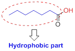
The structure of the micelle in a saturated fatty acid salt has to be drawn and their parts need to be shown.
Concept Introduction:
Micelle formation:
A sphere-shaped cluster formed when the accumulation of soap or detergent molecules so that their hydrophilic ends are on the surface and hydrophobic ends is in the centre are formed
Acids have two different parts
- (1) Hydrophilic (water-loving),
- (2) Hydrophobic (water-fearing).
Hydrophilic (water-loving):
The compound has the carboxyl group (

Hydrophobic (water-fearing):
The compound has alkyl chain group is called as hydrophilic.
Expert Solution & Answer
Want to see the full answer?
Check out a sample textbook solution
Students have asked these similar questions
Part a) Explain the entropic and enthalpic arguments for micelle formation.
Part b) Explain the entropic and enthalpic arguments for a bilayer formation.
Match the area of dipeptide with the number
α- D -Mannose is a sweettasting sugar. β - D -Mannose , on the other hand, tastes bitter. A pure solution of α - D -mannose loses its sweet taste with time as it is converted into the β anomer . Draw the β anomer and explain how it is formed from the α anomer
Chapter 23 Solutions
EBK FUNDAMENTALS OF GENERAL, ORGANIC, A
Ch. 23.1 - Use Figure 23.1 to identify the family of lipids...Ch. 23.2 - Prob. 23.2PCh. 23.2 - Prob. 23.3PCh. 23.2 - Prob. 23.4KCPCh. 23.3 - Prob. 23.1CIAPCh. 23.3 - Prob. 23.2CIAPCh. 23.3 - Prob. 23.3CIAPCh. 23.3 - Prob. 23.5PCh. 23.3 - Prob. 23.6PCh. 23.3 - Prob. 23.7KCP
Ch. 23.4 - Prob. 23.8PCh. 23.4 - Prob. 23.9PCh. 23.4 - Prob. 23.10PCh. 23.4 - Prob. 23.11PCh. 23.5 - Prob. 23.12PCh. 23.5 - Draw the structure of the sphingomyelin that...Ch. 23.5 - Draw the structure of the glycerophospholipid that...Ch. 23.5 - Prob. 23.16PCh. 23.7 - Prob. 23.17KCPCh. 23.7 - Prob. 23.4CIAPCh. 23.7 - Prob. 23.6CIAPCh. 23.7 - Prob. 23.7CIAPCh. 23.7 - Prob. 23.8CIAPCh. 23.7 - Prob. 23.18PCh. 23.7 - Prob. 23.19PCh. 23.7 - Prob. 23.20KCPCh. 23 - The fatty acid composition of three...Ch. 23 - Prob. 23.23UKCCh. 23 - According to the fluid-mosaic model (Figure 23.7),...Ch. 23 - Dipalmitoylphosphatidylcholine (DPPC) is a...Ch. 23 - Prob. 23.26APCh. 23 - Prob. 23.27APCh. 23 - Prob. 23.28APCh. 23 - Prob. 23.29APCh. 23 - Differentiate between saturated, monounsaturated,...Ch. 23 - Are the carboncarbon double bonds in naturally...Ch. 23 - Prob. 23.32APCh. 23 - Prob. 23.33APCh. 23 - Which of these fatty acids has the lower melting...Ch. 23 - Which of these fatty acids has the higher melting...Ch. 23 - Prob. 23.36APCh. 23 - Prob. 23.37APCh. 23 - Prob. 23.38APCh. 23 - Prob. 23.39APCh. 23 - What function does a wax serve in a plant or...Ch. 23 - Prob. 23.41APCh. 23 - Prob. 23.42APCh. 23 - What kind of lipid is spermacetia fat, a wax, or a...Ch. 23 - Prob. 23.44APCh. 23 - Prob. 23.45APCh. 23 - Prob. 23.46APCh. 23 - Prob. 23.47APCh. 23 - Prob. 23.48APCh. 23 - Prob. 23.50APCh. 23 - Prob. 23.52APCh. 23 - Prob. 23.53APCh. 23 - Describe the difference between a triacylglycerol...Ch. 23 - Why are glycerophospholipids, rather than...Ch. 23 - Prob. 23.56APCh. 23 - Prob. 23.57APCh. 23 - Why are glycerophospholipids more soluble in water...Ch. 23 - Prob. 23.59APCh. 23 - Prob. 23.60APCh. 23 - Prob. 23.61APCh. 23 - Draw the structure of a glycerophospholipid that...Ch. 23 - Prob. 23.63APCh. 23 - What is a major function of cholesterol in your...Ch. 23 - Prob. 23.65APCh. 23 - Prob. 23.66APCh. 23 - Prob. 23.67APCh. 23 - Explain how a micelle differs from a membrane...Ch. 23 - Prob. 23.69APCh. 23 - Prob. 23.70APCh. 23 - Prob. 23.71APCh. 23 - Prob. 23.72APCh. 23 - Prob. 23.73APCh. 23 - Prob. 23.74APCh. 23 - Prob. 23.75APCh. 23 - Draw the structure of a triacylglycerol made from...Ch. 23 - Prob. 23.79CPCh. 23 - Prob. 23.80CPCh. 23 - Explain why cholesterol is not saponifiable.Ch. 23 - Draw cholesterol acetate. Is this molecule...Ch. 23 - Prob. 23.83CPCh. 23 - Prob. 23.84CPCh. 23 - Prob. 23.85CPCh. 23 - Prob. 23.86CPCh. 23 - Prob. 23.88GP
Knowledge Booster


Learn more about
Need a deep-dive on the concept behind this application? Look no further. Learn more about this topic, biochemistry and related others by exploring similar questions and additional content below.Similar questions
- Draw the structure of ethanolamine sphingomyelin formed from linoleic acid Draw the structure of serine sphingomyelin formed from arachidic acidarrow_forwardThe following are structural diagrams of a selection of newly discovered amino acids. OH -の-CHs NH HO C-OH NH, AN-CH CH2 CH2 OH Ho NH, C=0 a) Select 1 amino acid. Redraw it. Label the alpha carbon and circle/highlight the entire backbone of the amino acid. b) The amino acids are part of a channel protein embedded in the cell membrane. Choose 2 amino acids (from above) that you would expect to find within the interior/middle of the cell membrane. Draw the formation of the dipeptide using the 2 amino acids you selected. Identify the other products formed in the reaction.arrow_forwardSoap molecules have a polar head and a non polar tail. Briefly explain whether your experimental observation of phycocyanin folding in the presence of soap solution is consistent with the structure of soap molecules. Results: almost colorless, clear and no fluorescencearrow_forward
- Draw the peptide formed between asparagine and histidine. H,N- -CH-C-OH H,N-CH-ċ-OH ČH2 ČH, N° NH2 -NH +arrow_forwardConsider the structure of the tripeptide below. H O 11 H₂N-C-C-N-C-c- CH₂ CH₂ C=0 NH₂ pH 5: Z-I pH 10: H HN H O 11 CH₂ NH HIC- I-Z 0=6 -N-C-C-OH 1. What is the sequence of the tripeptide? (use the one-letter symbol, do not put dash or space in between symbols) 2. What is the net charge of the dominant structure of the tripeptide at the given pH values? The pK, values of the amino acids are given in Table 1. CH-OH T CH3 Table 1. pk, values of the standard amino acids.arrow_forwardDraw the structural formula of the oligopeptide if the amino acids are arginine, glutamine, glycine, methionine and glutamic acid considering the first amino acid is the N-terminusarrow_forward
- A major ingredient in peanut butter cup candy is soy lecithin. Draw the structure of lecithin.arrow_forwardWhich structı Which structure is highlighted?arrow_forwardIn the figure below, protein 1 is located in the cytosol, and protein 2 is membrane bound. Give 3 specific examples from Figure 4-3 of amino acids that you might expect to find on the surface of protein 1. For protein 2, give three specific amino acids that you would expect to be on the surface near both points A and B (labeled with stars). To clarify, you should choose 3 amino acids for point A and also list three amino acids for point B. All of the amino acids you choose for Protein 2 must be different from those that you choose for Protein 1. Rationalize your choices by discussing the amino acids you chose, and their properties in a few sentences. Protein 1 Protein 2arrow_forward
- The lipid portion of a typical bilayer is about 3 nm thick. (a) Calculate the number of residues in an a helix that will just span this distance. (b) The epidermal growth factor receptor has a single transmem- brane helix. Find it in this partial sequence: .--RGPKIPSIATGMVGALLLLVVALGIGILFMRRRH-…arrow_forwardWater-soluble proteins such as myoglobin tend to fold such that: A) hydrophobic amino acids R-groups are on the interior of the protein and hydrophilic groups are on the outside OB) all peptides form hydrogen bonds with water. C) hydrophilic amino acid R-groups are on the interior of the protein and hydrophobic groups are on the outside. O D) hydrophilic and hydrophobic amino acid R-groups form hydrogen bonds with each other.arrow_forwardLabel: 1) the type of chemical bonds between the amino acids (e.g. covalent bond, ionic bond, metallic bond) 2) the type of interparticle forces of attraction occurring within the protein and with its environment *Indicate at least four observed interparticle forces of attraction *pink - negatively charged, blue - positively charged, yellow - nonpolar and uncharged, green - polar and uncharged *[See example picture] The chemical bond (shown by the arrow) is depicted as a line between the amino acids. Interparticle forces of attraction, such as the one between Phe and Glu (boxed), are not represented by lines but rather by the proximity of amino acids.arrow_forward
arrow_back_ios
SEE MORE QUESTIONS
arrow_forward_ios
Recommended textbooks for you
 BiochemistryBiochemistryISBN:9781319114671Author:Lubert Stryer, Jeremy M. Berg, John L. Tymoczko, Gregory J. Gatto Jr.Publisher:W. H. Freeman
 Lehninger Principles of BiochemistryBiochemistryISBN:9781464126116Author:David L. Nelson, Michael M. CoxPublisher:W. H. Freeman
 Fundamentals of Biochemistry: Life at the Molecul...BiochemistryISBN:9781118918401Author:Donald Voet, Judith G. Voet, Charlotte W. PrattPublisher:WILEY
 BiochemistryBiochemistryISBN:9781305961135Author:Mary K. Campbell, Shawn O. Farrell, Owen M. McDougalPublisher:Cengage Learning
 BiochemistryBiochemistryISBN:9781305577206Author:Reginald H. Garrett, Charles M. GrishamPublisher:Cengage Learning
 Fundamentals of General, Organic, and Biological ...BiochemistryISBN:9780134015187Author:John E. McMurry, David S. Ballantine, Carl A. Hoeger, Virginia E. PetersonPublisher:PEARSON
Biochemistry
Biochemistry
ISBN:9781319114671
Author:Lubert Stryer, Jeremy M. Berg, John L. Tymoczko, Gregory J. Gatto Jr.
Publisher:W. H. Freeman
Lehninger Principles of Biochemistry
Biochemistry
ISBN:9781464126116
Author:David L. Nelson, Michael M. Cox
Publisher:W. H. Freeman
Fundamentals of Biochemistry: Life at the Molecul...
Biochemistry
ISBN:9781118918401
Author:Donald Voet, Judith G. Voet, Charlotte W. Pratt
Publisher:WILEY
Biochemistry
Biochemistry
ISBN:9781305961135
Author:Mary K. Campbell, Shawn O. Farrell, Owen M. McDougal
Publisher:Cengage Learning
Biochemistry
Biochemistry
ISBN:9781305577206
Author:Reginald H. Garrett, Charles M. Grisham
Publisher:Cengage Learning
Fundamentals of General, Organic, and Biological ...
Biochemistry
ISBN:9780134015187
Author:John E. McMurry, David S. Ballantine, Carl A. Hoeger, Virginia E. Peterson
Publisher:PEARSON