
DRAW IT Below are the amino acid sequences(using the single-letter code; see Figure 5.14) of four short segments of the FOXP2 protein from six Speeles: chimpanzce (Q, orangutan (()), gorllla (G), rhesus macaque (R), mouse (M), and human (H). These segments contain all of the amino acid differences helween the FOXP2 proteinsof these species.
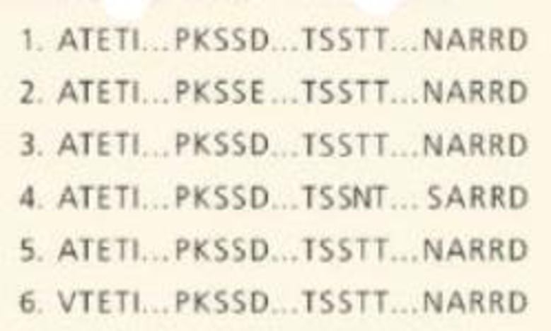
Use a highlighter to color anv amino acid that varies among the species. (Color that amino acid in all sequences.
- (a) The C, G, R sequences are identical. Identily which lines correspond to those sequences.
- (b) The O sequence differs from the C, G, R speieces at two amino acids. Underilnethetwodlffcrences inthe H sequence.
- (c) The O sequence diffen from the C, G, R sequences at one amino acid (having V instead of A) and from the H sequence at three amino acids. Identify tho O sequence.
- (d) In the M sequence,circle the amino acid(s) that differ from the C, G, R sequence, and draw a square around those that differ from the H sequence.
- (e) Primates and rodents diverged between 60 and IO0 million years ago. and chimpanzees and humans about 6 million years ago. Compare the amino acid differences between the mouse and the C, G, R species with those between the human and the C, G, R species. What can you conclude?

Learn your wayIncludes step-by-step video

Chapter 21 Solutions
Campbell Biology: Custom Edition
Additional Science Textbook Solutions
Human Biology: Concepts and Current Issues
Campbell Biology in Focus (2nd Edition)
Campbell Biology: Concepts & Connections (9th Edition)
Laboratory Experiments in Microbiology (12th Edition) (What's New in Microbiology)
Becker's World of the Cell (9th Edition)

- Construct a multiple protein sequence alignment using human HOXD13 and 9 other orthologues of human HOXD13. In the figure legend, describe what is being shown, and specify the size (start and stop number of amino acid) and location of conserved domain(s) in HOXD13 level: UNIarrow_forwardYou have discovered a novel protein that has a pl = 5.5. To study the functional properties of this new protein your research group has made a mutant that contains two amino acid changes-namely, a surface Phe residue in the normal protein has been replace by His (side chain pk = 6.1), and a surface Gln has been replace by Glu (side chain pk. = 6.0). The pl of the mutant protein is predicted to be: A. Greater than the pl of the normal protein. B. Less than the pl of the normal protein. C. The same as the pl of the normal protein.arrow_forwardIn a folded protein, Glu116 is close in three-dimensional space to Lys224. The pKa of the carboxylic acid functional group in the side chain of glutamic acid is 4.3. The pKa of the amino group in the side chain of lysine is 10.5. How do you think the pKa of the side chain of Glu116 will change in the folded protein? a. The pKa will be > 4.3. b. The pKa will be < 4.3. c. The pKa will not change. Use chemical reasoning to justify your answer.arrow_forward
- A pentapeptide was found to increase the rate of hydrolysis of fats in the adipose tissues to provide fatty acids as sources of energy. The amino acid sequence of this peptide was determined to be: ALA-ARG-LYS-MET-SER Hydrolysis of the three variants of the peptide, and analysis of their amino acid composition revealed the following changes: Variants Amino Acid Replacement Peptide 1 Arg was replaced by Leu Peptide 2 Ala was replaced by Tyr Peptide 3 Arg was replaced by Glu What will be the positions of these variant peptides compared with the normal peptide in an electrophoretic run at pH 7.0?arrow_forwardThe following steps were performed using enzyme cleavage of a peptide to determine its amino acid sequence. Step 1. FDNB yield DNB-Gly Step 2. Treatment with trypsin yield 3 fragments: Tyr-Leu-Asp-Arg; Gly-Ser-Ala-Lys; Trp-Gly-Ser-Met Step 3. Treatment with pepsin gave the same 3 peptide fragments. What is the sequence of the peptide?arrow_forwardAlthough the Shine-Dalgarno sequences vary considerably in differ- ent genes, they include examples like GAGGGG that could serve as code-in this case, for Glu-Gly. Does this imply that the sequence Glu-Gly cannot ever occur in a protein, lest it be read as a Shine-Dalgarno sequence? Speculate.arrow_forward
- Sequence: CCACCTGTACCCGGACACACCCTGGTGTCC What human disease has been connected to this gene? Calculate molecular weight (kiloDalton, kD) and calculated pI (the pH where the protein carries no net electrical charge) of the protein.arrow_forwardSickle cell anemia is caused by a point mutation in the β-globin chain of hemoglobin. Glutamic acid is replaced by Valine. HBB sequence in normal adult hemoglobin (Hb A): Leu-Thr-Pro-Glu-Glu-Lys-Ser HBB sequence in mutant adult hemoglobin (Hb S): Leu-Thr-Pro-Val-Glu-Lys-Ser What effect does this mutation have on the structure and function of the protein? Predict what would happen to the RBC if the glutamic acid was replaced with asparagine instead of valine.arrow_forwardA peptide with the primary structure Lys-Arg-Pro-Leu-Ile-Asp-Gly-Ala must be synthesized by the methods developed by Merrifield. Calculate the percentage of the peptides synthesized that will be full length and have the correct sequence if the addition of each amino acid residue is 96% efficient. Do the calculation a second time but assume a 99% efficiency for each cycle. full-length peptides with the correct sequence if 96% efficient: full-length peptides with the correct sequence if 99% efficient: % %arrow_forward
- Consider the following coding 71 nucleotide DNA template sequence (It does not contain a translational start): 5’- GTTTCCCCTATGCTTCATCACGAGGGCACTGACATGTGTAAACGAAATTCCAACCTGAGCGGCGT GTTGAG-3’ By in vitro translating the mRNA, you determined that the translated peptide is 15 amino acids long. What is the expected peptide sequence in single letter abbreviations?arrow_forwardA peptide with 19 amino acid residues was digested with trypsin and generated the following fragments: • GGIR SFLG WAAPK AEEGLR A similar peptide was treated with chymotrypsin and generated the following fragments: • LG AEEGLRW AAPKGGIRSF Elucidate the correct sequence of the peptide.arrow_forwardShown to the right is a cartoon image of ETC Complex I from Y. lipolytica. A group of authors studied this wild type Complex I and also a version where the arginine at position 121 in the chain 3 subunit was mutated to a methionine (R121M). Part of chain 3 subunit contains the peptide sequence SMITH a. Draw the titration curve for the SMITH peptide. Mark all pKa’s and the pI on your curve. For the SMITH peptide at physiological pH, explain what types of interactions you believe the side chains are or are not able to participate in.arrow_forward
 Human Anatomy & Physiology (11th Edition)BiologyISBN:9780134580999Author:Elaine N. Marieb, Katja N. HoehnPublisher:PEARSON
 Biology 2eBiologyISBN:9781947172517Author:Matthew Douglas, Jung Choi, Mary Ann ClarkPublisher:OpenStax
 Anatomy & PhysiologyBiologyISBN:9781259398629Author:McKinley, Michael P., O'loughlin, Valerie Dean, Bidle, Theresa StouterPublisher:Mcgraw Hill Education,
 Molecular Biology of the Cell (Sixth Edition)BiologyISBN:9780815344322Author:Bruce Alberts, Alexander D. Johnson, Julian Lewis, David Morgan, Martin Raff, Keith Roberts, Peter WalterPublisher:W. W. Norton & Company
 Laboratory Manual For Human Anatomy & PhysiologyBiologyISBN:9781260159363Author:Martin, Terry R., Prentice-craver, CynthiaPublisher:McGraw-Hill Publishing Co.
 Inquiry Into Life (16th Edition)BiologyISBN:9781260231700Author:Sylvia S. Mader, Michael WindelspechtPublisher:McGraw Hill Education