Study Guide for Campbell Biology
11th Edition
ISBN: 9780134443775
Author: Lisa A. Urry, Michael L. Cain, Steven A. Wasserman, Peter V. Minorsky, Jane B. Reece, Martha R. Taylor, Michael A. Pollock
Publisher: PEARSON
expand_more
expand_more
format_list_bulleted
Videos
Textbook Question
Chapter 49, Problem 4IQ
Identify the structures (a-g) in the following illustration of the human brain. Then match the functions (1-7) to these structures.
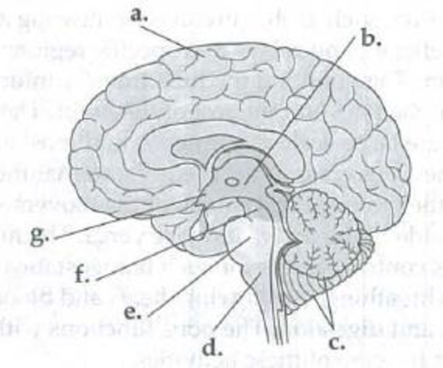
Functions
- 1. Coordinates balance and movement
- 2. Aids the medulla in some functions; conducts information between the brain and spinal cord
- 3. Sorts and relays information to the cerebrum
- 4. Regulates breathing, heart rate, and digestion
- 5. Integrates sensory and motor information; is center for learning, emotion, memory, and perception
- 6. Produces hormones; functions in homeostatic regulation
- 7. Sends sensory information to the forebrain; is involved in hearing and visual reflexes
Expert Solution & Answer
Want to see the full answer?
Check out a sample textbook solution
Students have asked these similar questions
Identify the parts of a neuron or a brain that correspond to the given functions
below. The parts are found inside the grid and loop these words either
horizontally, vertically, diagonally or inversely.
1. controls the growth of a nerve cell
2. carry impulses or messages toward the cell body
3. carry messages away from the cell body
4. serves as the body’s life support system
5. coordinates muscle movement
6. responsible for mental processes
7. controls breathing, heart rate and swallowing
8. controls the movement of the eye
9. regulates breathing and helps control eye movement
10. serves as a relay station for senses
Draw a lateral view of the human brain and label the major structures, including the lobes; write the functions of each lobe.
Mark the following statements about the brain as true or false. If a statement is false, correct it to make a true statement.a. Humans use only 10% of their brains.b. The four main components of the brain are the cerebrum, the diencephalon, the cerebellum, and the brainstem.c. The right and left lateral ventricles are the largest of the ventricles in the brain and are located in the diencephalon.d. The cerebrum is responsible for our basic, involuntary homeostatic functions and reflexes
Chapter 49 Solutions
Study Guide for Campbell Biology
Ch. 49 - The organization of an organisms nervous system...Ch. 49 - Prob. 2IQCh. 49 - Prob. 3IQCh. 49 - Identify the structures (a-g) in the following...Ch. 49 - Prob. 5IQCh. 49 - Action potentials are initiated more readily in...Ch. 49 - Prob. 1SYKCh. 49 - Prob. 1TYKCh. 49 - Prob. 2TYKCh. 49 - Prob. 3TYK
Knowledge Booster


Learn more about
Need a deep-dive on the concept behind this application? Look no further. Learn more about this topic, biology and related others by exploring similar questions and additional content below.Similar questions
- Match the function to the appropriate structure 1. Processes information about pain, pressure, and touch 2. controls and regulates heart rates, blood pressure, and breathing 3. Moves the muscles of the upper back 4. regulates the feeding reflex in infants 5. Adjust posture, maintains equilibrium, fine tunes movements 6. Moves the four eye muscles 7. Moves the tongue 8. Produces hormones such as ADH and oxytocin 9. relays and transmits sensory information to appropriate location in the brain 10. Collects taste information a. Midbrain b. trigeminal (Opthalmic and maxillary branches) c. Vagus nerve d. Accessory e. Hypothalamus f. Cerebellum g. Prefrontal association area h. Mammillary body i. Somatic sensory association area j. Oculomotor nerve k. Thalamus l. Somatic motor association area m. Gustatory cortex n. Hypoglossal o. Glossopharyngeal p. Facial q. Medulla Oblongata r. Pineal body s. Trochleararrow_forwardReview the functional regions of the cerebral cortex by matching each description with the appropriate letter in the figure below: 1. Receives information from cutaneous and musculoskeletal receptors 2. Interprets information from cutaneous and musculoskeletal receptors 3. Receives information from hair cells in the ears 4. Interprets information from hair cells in the ears 5. Plans skeletal movements 6. Issues commands that "tell" which muscles to contractarrow_forwardMark the following statements on the role of the brain in movement as true or false. If a statement is false, correct it to make a true statement. a. The dopaminergic neurons of the substantia nigra enhance the actions of the caudate nucleus and putamen. b. The cerebellum monitors the initiation of movement but does not monitor ongoing movements. c. The basal nuclei inhibit inappropriate movements and are required for the initiation of movement. d. The correction of motor error by the cerebellum can occur over the long term by motor learningarrow_forward
- Discuss the hierarchy of the human brain and the components that make up the hindbrain, midbrain, and forebrain. What is the function of each of these structures? Give real life examples of each component.arrow_forwardUnderstanding how your brain developed and what brain structures you share with other animals is crucial to understanding the bain and its functions. Describe the brain's formation, start with cell differentiation and end with central nervous system development (brain and spinal cord). The more detailed description the more you demonstrate your understanding. read:use your book and/or outside resourcesarrow_forwardThe indicated structure, behind the black arrow, joins lower parts of the ___________ and spinal cord with higher parts of the brain; includes nuclei for reflex centres. Fill in the blank, please!arrow_forward
- Which of the following brain regions is not correctly matched to itsfunction?a. The medulla oblongata regulates heartbeat, breathing, and bloodpressure.b. The cerebellum coordinates voluntary muscle movements.c. The thalamus secretes melatonin, which regulates daily bodyrhythms.d. The midbrain acts as a refl ex center for visual, auditory, andtactile responses.arrow_forwardWhich part of the human brain is the most developed?arrow_forwardWith a sketch and a line diagram show the development of the Brain from primary brain vesicle to adult structurearrow_forward
arrow_back_ios
SEE MORE QUESTIONS
arrow_forward_ios
Recommended textbooks for you

 Human Physiology: From Cells to Systems (MindTap ...BiologyISBN:9781285866932Author:Lauralee SherwoodPublisher:Cengage Learning
 Human Biology (MindTap Course List)BiologyISBN:9781305112100Author:Cecie Starr, Beverly McMillanPublisher:Cengage Learning
 Anatomy & PhysiologyBiologyISBN:9781938168130Author:Kelly A. Young, James A. Wise, Peter DeSaix, Dean H. Kruse, Brandon Poe, Eddie Johnson, Jody E. Johnson, Oksana Korol, J. Gordon Betts, Mark WomblePublisher:OpenStax College
Human Physiology: From Cells to Systems (MindTap ...
Biology
ISBN:9781285866932
Author:Lauralee Sherwood
Publisher:Cengage Learning
Human Biology (MindTap Course List)
Biology
ISBN:9781305112100
Author:Cecie Starr, Beverly McMillan
Publisher:Cengage Learning
Anatomy & Physiology
Biology
ISBN:9781938168130
Author:Kelly A. Young, James A. Wise, Peter DeSaix, Dean H. Kruse, Brandon Poe, Eddie Johnson, Jody E. Johnson, Oksana Korol, J. Gordon Betts, Mark Womble
Publisher:OpenStax College
Embryology | Fertilization, Cleavage, Blastulation; Author: Ninja Nerd;https://www.youtube.com/watch?v=8-KF0rnhKTU;License: Standard YouTube License, CC-BY