
Videos
In an experiment published in the journal Cell in 2014, Amnon Koren and Steven McCarroll isolated two populations of growing tissue culture cells from each of two unrelated people from different parts of the world.
One population from each person consisted of millions of cells that were in G of the cell cycle; the other population was a similar number of cells that were in S phase for various amounts of time. The scientists then performed high-throughput DNA sequencing on these cell populations.
The two graphs that follow show the data for the two individuals. In each graph, the x-axis represents positions along a chromosome (here, chromosome 8), and the y-axis represents the ratio between the number of reads obtained for a given region of the genome from the S phase sample divided by the number of reads obtained for the same region from the G sample. Each small purple dot is 2 kb along the chromosome; the black line is the moving average of the purple.
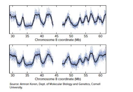
| a. | At chromosomal coordinate 33 Mb, the y-axis value is much higher than at coordinate 35 Mb. What does this fact tell you about the timing of |
| b. | Scientists still do not have a good idea about the nature of DNA sequences or chromatin structures that define origins of replication in human cells. If you were trying to locate such origins of replication, where would you look? |
| c. | Suppose you did a similar experiment using two populations with the same number of cells, one population in G and the other in G . If you graphed the data in a similar fashion, with the y-axis representing the ratio of the number of reads from the G sample divided by the number of reads from the G sample, what would the plot look like? |
| d. | The patterns for these two people are very nearly the same, even though they are completely unrelated. What does this fact suggest? |
| e. | These scientists later reasoned that they could obtain the same kind of information from any person whose genome had been sequenced by high-throughput methods, without separating out populations of cells at different cell cycle stages. What would have to be true about the cells analyzed and the kinds of data available? Why would you want to look at this data from many different people? |
Want to see the full answer?
Check out a sample textbook solution
Chapter 12 Solutions
ND STONY BROOK UNIVERSITY LOOSELEAF GENETICS: FROM GENES TO GENOMES


- What have scientists discovered about the limiting factors that control how many times a cell can replicate itself and divide? Question 10 options: a) Telomeres do not play a role as a limiting factor. b) Telomeres get longer the more times a cell has replicated and divided. c) When telomeres become too short (or disappear), a cell stops dividing. d) None of the abovearrow_forwardSuppose you isolated a mutant strain of yeast that replicates its DNA more than once per cell cycle. In other words, each gene in the genome was replicated several times between successive cell divisions. How might you explain such a phenomenon?arrow_forwardTo identify genes controlling the cell cycle in budding yeast, a genetic screen was carried out. In this screen, haploid yeast cells were exposed to a DNA damaging agent to introduce random mutations in the genome. By culturing cells at an elevated temperature (e.g. 37 degrees), where many mutated genes lose their function, scientists identified yeast mutants that showed growth defects and arrest at specific stages of the cell cycle (e.g. in mitosis with large buds). In this screen, mutants of the cyclin-dependent kinase were identified, but not mutants of cyclins. Explain the reason for this outcome.arrow_forward
- Do a few cells created by therapeutic cloning of your own somatic cells constitute life? If these cells do constitute life, do they have the same rights as a human being conceived naturally? If it were possible, should someone be allowed to grow his or her own therapeutic clone into an adult?arrow_forward1) Scientists isolate cells in various phases of the cell cycle. They isolate a group of cells that have 1.5 times more DNA than G1 phase cells. What is the most likely part of the cell cycle from which these cells were isolated? A) in the G2 phase of the cell cycle. B) between the G1 and S phases in the cell cycle C) in the late M phase of the cell cycle D) in the S phase of the cell cycle 2) Which of the following statements is true regarding animal cell cytokinesis when compared to plant cell cytokinesis? A) The contractile filaments found in plant cells are structures composed of carbohydrates; the cleavage furrow in animal cells is composed of contractile proteins. B) Plant cells divide after metaphase but before anaphase; animal cells divide after anaphase. C) Animal cells form a cleavage furrow while plant cells deposit vesicles containing cell wall building blocks on the metaphase plate D) In animal cells, a cell membrane separates the two daughter cells; the structural…arrow_forwardThe microscope image above shows the human chromosomes from a white blood cell. To create the image, researchers put cells in culture under conditions that encourage the cells to divide. They bathed the cells in a hypotonic (low salt) solution, which caused the cells to swell until their plasma membrane burst open. They "squashed" the chromosomes to spread them out, and stained them with a dye to make them visible under the microscope. Human chromosomes are numbered from longest (1) to shortest (22) plus the sex chromosomes X and Y. In the image chromosome 1 is about 7 micrometers. Answer the following questions. 1) What word(s) in the description above indicates that the chromosomes are not from a cell undergoing meiosis? 2) Based on the size, shape and appearance of the chromosomes in the image, in what cell cycle stage was the cell that the chromosomes came from? How can you tell? 3) Does the image suggest that centromere sequences are always located in the middle of a…arrow_forward
- In an experiment to investigate membrane integrity, beet root sections were subjected to different chemicals. Membrane damage was estimated by the color intensity of leaked pigment. Five replicates of each treatment were prepared. Complete the following table with the absorbance values. Calculate the mean (average) and the standard Table 1: Absorbance readings of pigment leaked from damaged cells treated with two different chemicals. Replicate # Absorbance at 460 nm Chemical 2 Chemical 6 1 0.357 0.392 2 0.576 0.395 3 0.334 0.385 4 0.197 0.399 5 0.334 0.403 Average SD Calculate the Coefficient of Variance (see the appendix) for the readings of each chemical: Evaluate the precision of each and state which one is more precise:arrow_forwardThe technique of fluorescence in situ hybridization (FISH) is described. This is another method for examining sequence complexity within a genome. In this method, a DNA sequence, such as a particular gene sequence, can be detected within an intact chromosome by using a DNA probe that is complementary to the sequence.For example, let’s consider the β-globin gene, which isfound on human chromosome 11. A probe complementary to theβ-globin gene binds to that gene and shows up as a brightly colored spot on human chromosome 11. In this way, researchers can detectwhere the β-globin gene is located within a set of chromosomes. Becausethe β-globin gene is unique and because human cells are diploid(i.e., have two copies of each chromosome), a FISH experimentshows two bright spots per cell; the probe binds to each copy ofchromosome 11. What would you expect to see if you used thefollowing types of probes?A. A probe complementary to the Alu sequenceB. A probe complementary to a tandem array near…arrow_forwardSuppose one were provided with an actively dividing culture of E. coli to which radioactive thymine had been added. What would happen if a cell replicated once in the presence of the radioactive base? (a) one of the daughter cells, but not the other would have radioactive DNA (b) neither of the two daughter cells would be radioactive (c) only the purines of the DNA would be radioactive (d) radioactive thymine would pair with radioactive guanine (e ) DNA in both daughter cells would be radioactivearrow_forward
- One way that researchers create a cell line that is "immortal" (divides forever in culture) is by introducing DNA that forces the cell to express telomerase. Why do cells that don't express the telomerase protein stop dividing? What specifically goes wrong?arrow_forwardWhich of the following is TRUE regarding the Steward's (1950s) carrot experiment and Gurdon et al. (1975) nuclear transplantation experiment? O While both studies supported the differential expression of genetic material hypothesis, Gurdon et al. (1975) highlighted the role of differentiation on the genetic potential of a nucleus. O While both studies supported the "progressive loss of genetic material hypothesis, Gurdon et al. (1975) highlighted the role of differentiation on the genetic potential of a nucleus. O While Gurdon et. al's (1975) study clearly supported the differential expression" hypothesis, Steward's (1950s) carrots supported the progressive loss" hypothesis, highlighting the role of differentiation on nuclear potential. O While Steward's (1950s) carrot experiment clearly supported the "differential expression hypothesis, Gurdon et. al's (1975) study supported the "progressive lass" hypothesis, highlighting the role of differentiation on nuclear potential.arrow_forwardI am trying to understand DNA/chromosome structure and replication. I have a culture dish of human cells (NIH3T3) and expose them to radioactively labeled Thymidine, 3H-thymidine, during S phase of the cell cycle. How would my radioactivity be distributed over a pair of homologous chromosomes at metaphase? If i have to draw How the radioactivity would be distributed over a pair of homologous chromosomes at metaphase, how will my drwing look likearrow_forward
 Biology: The Dynamic Science (MindTap Course List)BiologyISBN:9781305389892Author:Peter J. Russell, Paul E. Hertz, Beverly McMillanPublisher:Cengage Learning