Campbell Biology, Books a la Carte Edition (11th Edition)
11th Edition
ISBN: 9780134154121
Author: Lisa A. Urry, Michael L. Cain, Steven A. Wasserman, Peter V. Minorsky, Jane B. Reece
Publisher: PEARSON
expand_more
expand_more
format_list_bulleted
Concept explainers
Videos
Textbook Question
Chapter 6, Problem 10TYU
SYNTHESIZE YOUR KNOWLEDGE
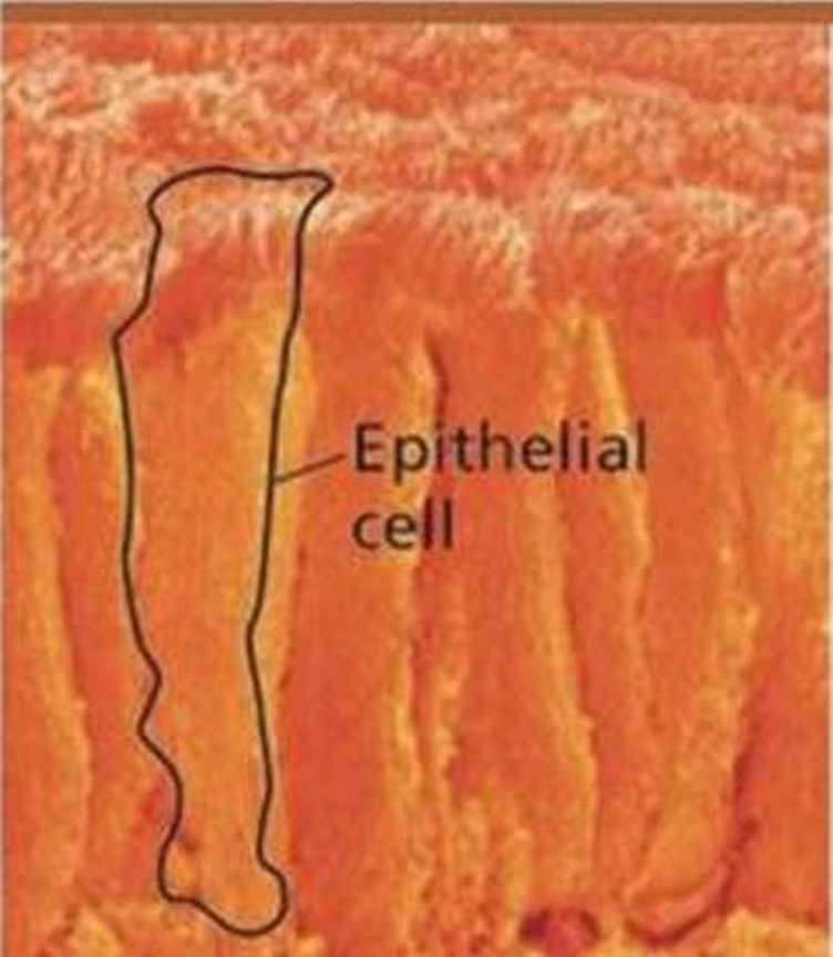
The cells in this SEM are epithelial cells from the small intestine. Discuss how aspects of their structure contribute to their specialized functions of nutrient absorption and as a barrier between the intestinal contents and the blood supply on the other side of the sheet of epithelial cells
.
Expert Solution & Answer
Want to see the full answer?
Check out a sample textbook solution
Students have asked these similar questions
Name the 4 different cell-cell contacts that are important for eg the function of the intestinal epithelium
Explain in general how each individual cell-cell contact works and how they contribute to the epithelium's resistance to pressure / tensile forces
Draw a plan diagram of the frog intestine viewed under the microscope (see attached images for help!!)....
YOU NEED TO DO A DRAWING AND DO CALCULATIONS. DO NOT ANSWER THIS WITH A WRITTEN RESPOSE!!!!
A plan diagram is a simple drawing showing only the boundaries between the lumen, the columnar epithelium, the (areolar) connective tissue, the circular smooth muscle and the longitudinal smooth muscle. A plan diagram serves to illustrate the location and relative thickness of the various tissue layers.
1. Draw a plan diagram and label the following: • Longitudinal smooth muscle • Columnar epithelial tissue • Circular smooth muscle • Villi • Areolar connective tissue • Lumen
2. Calculate actual size and drawing magnification of the width of the intestine. Include formulae used and calculations.
3. Add a scale bar with actual size next to your diagram.
Give your drawing a descriptive title and record total magnification.
Attached are an example plan diagram labelled, as well as a…
The smooth muscle cells are arranged in two different ways in intestinal wall. Explain the significance of two different arrangements of smooth muscle cells in intestinal wall i.e. how these arrangements relate to the function.
Chapter 6 Solutions
Campbell Biology, Books a la Carte Edition (11th Edition)
Ch. 6.1 - Prob. 1CCCh. 6.1 - Prob. 2CCCh. 6.2 - Briefly describe the structure and function of the...Ch. 6.2 - DRAW IT Draw a simplified elongated cell that...Ch. 6.3 - What role do ribosomes play in carrying out...Ch. 6.3 - Describe the molecular composition of nucleoli and...Ch. 6.3 - Prob. 3CCCh. 6.4 - Describe the structural and functional...Ch. 6.4 - Describe how transport vesicles integrate the...Ch. 6.4 - WHAT IF? Imagine a protein that functions in the...
Ch. 6.5 - Describe two characteristics shared by...Ch. 6.5 - Prob. 2CCCh. 6.5 - Prob. 3CCCh. 6.6 - Prob. 1CCCh. 6.6 - WHAT IF? Males afflicted with Kartagener's...Ch. 6.7 - In what way are the cells of plants and animals...Ch. 6.7 - Prob. 2CCCh. 6.7 - MAKE CONNECTIONS The polypeptide chain that makes...Ch. 6.8 - Prob. 1CCCh. 6 - Prob. 6.1CRCh. 6 - Explain how the compartmental organization of a...Ch. 6 - Describe the relationship between the nucleus and...Ch. 6 - Describe the key role played by transport vesicles...Ch. 6 - What does the endosymbiont theory propose us the...Ch. 6 - Describe the role of motor proteins inside the...Ch. 6 - Prob. 6.7CRCh. 6 - When a cell ingests a bacterium, what role does...Ch. 6 - Which structure is not part of the endomembrane...Ch. 6 - Prob. 2TYUCh. 6 - Which of the following is present in a prokaryotic...Ch. 6 - Prob. 4TYUCh. 6 - Which cell would be best for studying lysosomes?...Ch. 6 - Prob. 6TYUCh. 6 - EVOLUTION CONNECTION (a) What cell structures best...Ch. 6 - SCIENTIFIC INQUIRY Imagine protein X, destined to...Ch. 6 - WRITE ABOUT A THEME: ORGANIZATION Considering some...Ch. 6 - SYNTHESIZE YOUR KNOWLEDGE The cells in this SEM...
Additional Science Textbook Solutions
Find more solutions based on key concepts
Explain why hyperthermophiles do not cause disease in humans.
Microbiology with Diseases by Taxonomy (5th Edition)
A student moving out of a dormitory crouches in correct fashion to lift a heavy box of books. What prime movers...
HUMAN ANATOMY
What are the cervical and lumbar enlargements?
Principles of Anatomy and Physiology
Some people compare DNA to a blueprint stored in the office of a construction company. Explain how this analogy...
Biology: Concepts and Investigations
Knowledge Booster


Learn more about
Need a deep-dive on the concept behind this application? Look no further. Learn more about this topic, biology and related others by exploring similar questions and additional content below.Similar questions
- Select the best answer or answers from the choices given: A pancreas cell makes proteins (enzymes) that it releases to the small intestine. Which of the following best describes the path of these proteins from synthesis to exocytosis at the pancreatic cell’s plasma membrane (PM)? (a) Golgi S rough ER S PM, (b) smooth ER S Golgi S lysosome S PM, (c) rough ER S Golgi S PM, (d) nucleus S Golgi S PM.arrow_forwardDraw a plan diagram of the frog intestine viewed under the microscope (see attached images for help!!).... A plan diagram is a simple drawing showing only the boundaries between the lumen, the columnar epithelium, the (areolar) connective tissue, the circular smooth muscle and the longitudinal smooth muscle. A plan diagram serves to illustrate the location and relative thickness of the various tissue layers. 1. Draw a plan diagram and label the following: • Longitudinal smooth muscle • Columnar epithelial tissue • Circular smooth muscle • Villi • Areolar connective tissue • Lumen 2. Calculate actual size and drawing magnification of the width of the intestine. Include formulae used and calculations. 3. Add a scale bar with actual size next to your diagram. Give your drawing a descriptive title and record total magnification. Attached are an example plan diagram labelled, as well as a microscopic image of the frog intestine (3-4 cells should fit across only) PLEASE DO THE DRAWING AND…arrow_forwardObjective: Drawing a plan diagram of a frog intensine viewed under a microscope, as well as calculating actual size and drawing magnification of the width of the intestine. Please see the attached microscopic image of the frog intestine when viewed under the microscope, and an example labeled drawing of the intestine. I approximated that 3-4 cells would fit across (frog intenstine is much smaller when viewed under microscope, photo taken appears much longer). 1. Draw a plan diagram and label the following: • Longitudinal smooth muscle • Columnar epithelial tissue • Circular smooth muscle • Villi • Areolar connective tissue • Lumen 2. Calculate actual size and drawing magnification of the width of the intestine. Include formula used and calculations. 2. Add a scale bar with actual size next to the diagram. 3. Give the drawing a descriptive title and record total magnification. Thank you!arrow_forward
- The steps of how to make a wet mouth of cheek epithelial cells.place the steps of how to prepare a wet mouth in the correct order form left to right?arrow_forwardWhich of the following is NOT true about the peroxisome? It is _____. a. assembles from proteins b. a membrane-bound organelle c. responsible for packaging proteins d. similar to the lysosomearrow_forwardA virus can easily manipulate various cellular activities once it has already invadedthe host cell. Does this mean that a virus is a living organism because it can enter acell, the fundamental unit of life? Provide a brief explanation as to its relationship with the principle of cell theoryinvolved. Thank youarrow_forward
- Objective: Drawing a plan diagram of a frog intensine viewed under a microscope, as well as calculating actual size and drawing magnification of the width of the intestine. Please see the attached microscopic image of the frog intestine when viewed under the microscope, and an example labeled drawing of the intestine. I approximated that 3-4 cells would fit across (frog intenstine is much smaller when viewed under microscope, photo taken appears much longer). DO THE FOLLOWING BELOW STEPS: You need to complete a drawing. Please don't submit wrong answer. 1. Draw a plan diagram and label the following: • Longitudinal smooth muscle • Columnar epithelial tissue • Circular smooth muscle • Villi • Areolar connective tissue • Lumen 2. Calculate actual size and drawing magnification of the width of the intestine. Include formula used and calculations. 2. Add a scale bar with actual size next to the diagram. 3. Give the drawing a descriptive title and record total magnification.arrow_forwardHow are secretory proteins built, modified, and transported to the outer cell membrane. Make sure to mention the following organelles or cell parts in your answer: cell membrane, rough endoplasmic reticulum, Golgi, nucleus, ribosome, vesicles.arrow_forwardA human pancreatic cell obtains O2—and necessary molecules such as glucose, amino acids, and cholesterol—from its environment, and it releases CO2 as a waste product. In response to hormonal signals, the cell secretes digestive enzymes. It also regulates its ion concentrations by exchange with its environment. Based on the structure and function of cellular membranes, describe how such a cell accomplishes these interactions with its environment.arrow_forward
- Which of the following is not a part of animal cells' ECM? Select one: a. cellulose b. elastin c. Proteoglycans such as glycosaminoglycan (GAGs) d. Collagen e. Fibronectinarrow_forwardWhen William H. was helping victims after a devastating earthquake in a region not prepared to swiftly set up adequate temporary shelter, he developed severe diarrhea. He was diagnosed as having cholera, a disease transmitted through unsanitary water supplies contaminated by fecal material from infected indiv iduals. The toxin produced by cholera bacteria causes Cl- channels in the lurninal membranes of the intestinal cells to stay open, thereby increasing the secretion of Cl- from the cells into the intestinal tract lumen. By what mechanisms would Na+ and water be secreted into the lumen in conjunction with Cl- secretion? How does this secretory response account for the severe diarrhea that is characteristic of cholera?arrow_forwardTwo students are designing 3D cell models for animal cells.what organelles will the student need in order to build an accurate representation of an animal cell?arrow_forward
arrow_back_ios
SEE MORE QUESTIONS
arrow_forward_ios
Recommended textbooks for you
 Anatomy & PhysiologyBiologyISBN:9781938168130Author:Kelly A. Young, James A. Wise, Peter DeSaix, Dean H. Kruse, Brandon Poe, Eddie Johnson, Jody E. Johnson, Oksana Korol, J. Gordon Betts, Mark WomblePublisher:OpenStax College
 Biology (MindTap Course List)BiologyISBN:9781337392938Author:Eldra Solomon, Charles Martin, Diana W. Martin, Linda R. BergPublisher:Cengage Learning
 Human Physiology: From Cells to Systems (MindTap ...BiologyISBN:9781285866932Author:Lauralee SherwoodPublisher:Cengage Learning
Anatomy & Physiology
Biology
ISBN:9781938168130
Author:Kelly A. Young, James A. Wise, Peter DeSaix, Dean H. Kruse, Brandon Poe, Eddie Johnson, Jody E. Johnson, Oksana Korol, J. Gordon Betts, Mark Womble
Publisher:OpenStax College
Biology (MindTap Course List)
Biology
ISBN:9781337392938
Author:Eldra Solomon, Charles Martin, Diana W. Martin, Linda R. Berg
Publisher:Cengage Learning
Human Physiology: From Cells to Systems (MindTap ...
Biology
ISBN:9781285866932
Author:Lauralee Sherwood
Publisher:Cengage Learning
Types of Human Body Tissue; Author: MooMooMath and Science;https://www.youtube.com/watch?v=O0ZvbPak4ck;License: Standard YouTube License, CC-BY