
Videos
HeLa Cells Are a Genetic Mess HeLa cells can vary in chromosome number. Defects in proteins that orchestrate cell division result in descendant cells with too many or too few chromosomes, an outcome that is one of the ha1lmarks of cancer cells. The panel of chromosomes in FIGURE 11.9, originally published in 1989, shows all of the chromosomes in a single metaphase HeLa cell.
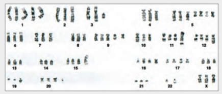
FIGURE 11.9 Karyotype of HeLa showing chromosomes in one cell.
What is the chromosome number of this HeLa cell?
To determine: The number of chromosomes in the HeLa cell.
Concept introduction: HeLa cell is the oldest and most commonly used cell line that was derived from the cervical cancer cells. HeLa cells differ from the normal cells in many ways. These cells have the ability to contaminate other cell lines. These cells have furthered the understanding of cancer, HIV, and the cells in general. It is still used widely to grow viruses and to test anti-tumor medicines.
Answer to Problem 1DAA
Explanation of Solution
Want to see more full solutions like this?
Chapter 11 Solutions
Biology: The Unity and Diversity of Life - With MindTap
Additional Science Textbook Solutions
Human Anatomy (8th Edition)
Campbell Biology: Concepts & Connections (8th Edition)
Seeley's Anatomy & Physiology
Biology: Concepts and Investigations


- HeLa Cells Are a Genetic Mess HeLa cells can vary in chromosome number. Defects in proteins that orchestrate cell division result in descendant cells with too many or too few chromosomes, an outcome that is one of the ha1lmarks of cancer cells. The panel of chromosomes in FIGURE 11.9, originally published in 1989, shows all of the chromosomes in a single metaphase HeLa cell. FIGURE 11.9 Karyotype of HeLa showing chromosomes in one cell. How many extra chromosomes does this cell have, compared to a normal human body cell?arrow_forwardHeLa Cells Are a Genetic Mess HeLa cells can vary in chromosome number. Defects in proteins that orchestrate cell division result in descendant cells with too many or too few chromosomes, an outcome that is one of the ha1lmarks of cancer cells. The panel of chromosomes in FIGURE 11.9, originally published in 1989, shows all of the chromosomes in a single metaphase HeLa cell. FIGURE 11.9 Karyotype of HeLa showing chromosomes in one cell. Can you tell that this cell came from a female? How?arrow_forwardHeLa Cells Are a Genetic Mess HeLa cells can vary in chromosome number. Defects in proteins that orchestrate cell division result in descendant cells with too many or too few chromosomes, an outcome that is one of the ha1lmarks of cancer cells. The panel of chromosomes in FIGURE 11.9, originally published in 1989, shows all of the chromosomes in a single metaphase HeLa cell. FIGURE 11.9 Karyotype of HeLa showing chromosomes in one cell. How many extra chromosomes does this cell have, compared to a normal human body cell?arrow_forward
- After mitosis, each daughter cell contains genetic instructions that are ______ and _____ chromosome number of the parent cell. a. identical to the parent cells; the same b. identical to the parent cells; one-half the c. rearranged; the same d. rearranged; one-half thearrow_forwardHeLa Cells Are a Genetic Mess HeLa cells can vary in chromosome number. Defects in proteins that orchestrate cell division result in descendant cells with too many or too few chromosomes, an outcome that is one of the ha1lmarks of cancer cells. The panel of chromosomes in FIGURE 11.9, originally published in 1989, shows all of the chromosomes in a single metaphase HeLa cell. FIGURE 11.9 Karyotype of HeLa showing chromosomes in one cell. Can you tell that this cell came from a female? How?arrow_forwardYOU DON'T NEED TO EXPLAIN THESE QUESTIONS JUST PROVIDE ANSWER DNA separates during ____ of mitosis. a)Prophase b)Telophase c)Metaphase d)Interphase e)Anaphase 2. What separates during mitosis? a)Single DNA strands b)Cytoplasm c)Sister chromatids d)Telomeres e)Homologous chromosomes 3. Oncogenes are associated with cancer because they Oncogenes are associated with cancer because they (select one answer) Cause cells to initiate a death pathway Fail to put a “brake” on the cell cycle Push cells through the cell cycle Divert cells into G0 phase Slow down the cell cyclearrow_forward
- Figure 6.4 Which of the following is the correct order of events in mitosis?a. Sister chromatids line up at the metaphase plate. The kinetochore becomes attached to the mitotic spindle. The nucleus re-forms and the cell divides. The sister chromatids separate.b. The kinetochore becomes attached to the mitotic spindle. The sister chromatids separate. Sister chromatids line up at the metaphase plate. The nucleus re-forms and the cell divides.c. The kinetochore becomes attached to metaphase plate. Sister chromatids line up at the metaphase plate. The kinetochore breaks down and the sister chromatids separate. The nucleus re-forms and the cell divides.d. The kinetochore becomes attached to the mitotic spindle. Sister chromatids line up at the metaphase plate. The kinetochore breaks apart and the sister chromatids separate. The nucleus re-forms and the cell divides.arrow_forwardPlease help Place the images of the cell division in the right order and label them a)  What is the final product of this type of cell division? Indicate the number of dauahter cells, the TYPE OF CELLS (somatic cells? sex cells? other?), where in the body this process takes place, whether they are genetically diverse pridentical, haploid or diploid, the chromosome number in human cells, whether they contain sinale- or double-stranded chromosomes, and what the "fate" of these cells is i.e. what will they go on to do, if given the chance)?arrow_forwardHumans have ___ matching chromosomes. A human cell contains these ___ chromosomes because we go through a process called ___________ reproduction. In this process gametes are made which each contain only ___ of each chromosome. Then during ___________, a zygote receives one of each chromosome: ___ from Dad’s sperm & ___ from Mom’s egg. This is what restores the __________ number of chromosomes (46 for humans) that are necessary for life to continue.arrow_forward
- The following diagram shows a simulated microscopic view of a tissue sample taken from a patient where the cells have been squashed onto a slide and stained to visualize the cell's DNA. In the diagram, click or tap on the center of all cells that appear to be involved in any of the four stages of mitosis. Make sure to mark as close to the center of the cell as possible. In the diagram from Question 5, count the total number of cells visible and write the total number of cells in the space below: In the diagram from Question 5, count the number of cells that you had marked as currently undergoing any of the four phases of mitosis. How many cells in total are currently undergoing mitosis? Write this number in the space below: The mitotic index is a calculated value that represents the percentage of cells in a sample that are actively dividing. It involves counting the total number of cells present including those actively dividing or those in interphase, the total number of cells that…arrow_forwardRose plants are octoploid (octo = 8). Gametes from a rose plant contain 40 chromosomes. Indicate which of the following are TRUE statements. Select 3 correct answer(s) Question 11 options: The gametes from a rose plant are diploid. The basic chromosome number of a rose plant cell is 40. The number of chromatids in a rose plant cell at G2 of the cell cycle is 160. The number of chromatids in a rose plant cell at G2 of the cell cycle is 80. The gametes from a rose plant are tetraploid. The basic chromosome number of a rose plant cell is 10. Rose plants are aneuploid.arrow_forwardThrough a microscope, you can see a cell plate beginning to develop across the middle of a cell and nuclei forming on either side of the cell plate. This cell is most likely? If the DNA content of a DIPLOID cell in the G1 phase of the cell cycle is represented by 'x. then the DNA content of the same cell at metaphase of MElosIs I would be? 3.Cell A has half as much DNA as cells B. C. and D in a mitotically active tissue. Cell A is likelv in? 4. Which of the following is a recessive trait © Polydactyly © Albinism © Huntington Disease Juvenile glaucoma O None are correctarrow_forward
 Biology: The Unity and Diversity of Life (MindTap...BiologyISBN:9781337408332Author:Cecie Starr, Ralph Taggart, Christine Evers, Lisa StarrPublisher:Cengage Learning
 Biology: The Unity and Diversity of Life (MindTap...BiologyISBN:9781305073951Author:Cecie Starr, Ralph Taggart, Christine Evers, Lisa StarrPublisher:Cengage Learning
 Biology Today and Tomorrow without Physiology (Mi...BiologyISBN:9781305117396Author:Cecie Starr, Christine Evers, Lisa StarrPublisher:Cengage Learning
 Human Biology (MindTap Course List)BiologyISBN:9781305112100Author:Cecie Starr, Beverly McMillanPublisher:Cengage Learning