
Concept explainers
Videos
Bacteriophage-Inspired Antibiotics Although bacteriophages have been infecting bacteria for billions of years, no mechanism, has evolved in bacteria to prevent the viruses from lysing the cell walls of their hosts. Now, scientists are targeting the same bacterial wall components that bacteriophages do. The goal is to develop antibiotics that bacteria will be less likely to develop resistance to.
FIGURE 20.22 shows the results of a study to test Epimerox, a new bacteriophage-inspired antibiotic, against Bacillus anthracis, the bacterial species that causes the disease anthrax.
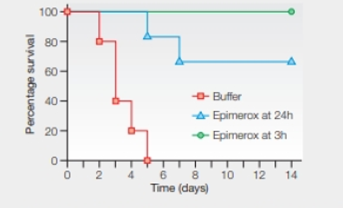
FIGURE 20.22 Effect of Epimerox on the survival of mice with anthrax. Mice were infected with the bacteria B. anthracis. One group of 15 then began receiving a drug-free buffer solution 3 hours later. Another 15 were treated with Epimerox beginning 3 hours after infection. A third group of 15was treated with Epimerox beginning 24 hours after infection.
In studies with Bacillus anthracis cells grown in culture, no Epimerox-resistant cells were observed. Explain why this result is consistent with the scientists' goal for developing this drug.
Want to see the full answer?
Check out a sample textbook solution
Chapter 20 Solutions
Biology: The Unity and Diversity of Life (MindTap Course List)
Additional Science Textbook Solutions
Human Anatomy
Microbiology with Diseases by Taxonomy (5th Edition)
Human Physiology: An Integrated Approach (7th Edition)
Campbell Essential Biology with Physiology (5th Edition)
HUMAN ANATOMY
Microbiology Fundamentals: A Clinical Approach - Standalone book


- HersheyChase Experiments The graph shown in FIGURE 8.5 is reproduced from an original 1952 publication by Hershey and Chase. Bacteriophage were labeled with radioactive tracers and allowed 10 infect bacteria. The virusbacteria mixtures were then whirled in a blender to dislodge any viral components attached to the exterior of the bacteria. Afterward, radioactivity from the tracers was measured. FIGURE 8.5 Detail of Alfred Hershey and Martha Chases 1952 publication describing their experiments with bacteriophage. Infected bacteria refers to the percentage of bacteria that survived the blender. After 4 minutes in the blender, what percentage of each isotope was extracellular?arrow_forwardHersheyChase Experiments The graph shown in FIGURE 8.5 is reproduced from an original 1952 publication by Hershey and Chase. Bacteriophage were labeled with radioactive tracers and allowed 10 infect bacteria. The virusbacteria mixtures were then whirled in a blender to dislodge any viral components attached to the exterior of the bacteria. Afterward, radioactivity from the tracers was measured. FIGURE 8.5 Detail of Alfred Hershey and Martha Chases 1952 publication describing their experiments with bacteriophage. Infected bacteria refers to the percentage of bacteria that survived the blender. The extracellular concentration of which isotope increased the most with blending?arrow_forwardHersheyChase Experiments The graph shown in FIGURE 8.5 is reproduced from an original 1952 publication by Hershey and Chase. Bacteriophage were labeled with radioactive tracers and allowed 10 infect bacteria. The virusbacteria mixtures were then whirled in a blender to dislodge any viral components attached to the exterior of the bacteria. Afterward, radioactivity from the tracers was measured. FIGURE 8.5 Detail of Alfred Hershey and Martha Chases 1952 publication describing their experiments with bacteriophage. Infected bacteria refers to the percentage of bacteria that survived the blender. Before blending what percentage of each isotope. 35S and 32P, was extracellular (outside the bacteria)?arrow_forward
- HersheyChase Experiments The graph shown in FIGURE 8.5 is reproduced from an original 1952 publication by Hershey and Chase. Bacteriophage were labeled with radioactive tracers and allowed 10 infect bacteria. The virusbacteria mixtures were then whirled in a blender to dislodge any viral components attached to the exterior of the bacteria. Afterward, radioactivity from the tracers was measured. FIGURE 8.5 Detail of Alfred Hershey and Martha Chases 1952 publication describing their experiments with bacteriophage. Infected bacteria refers to the percentage of bacteria that survived the blender. Do these results imply that viruses inject DNA or protein into bacteria? Why or why not?arrow_forwardFill in the blanks. The parentheses after each blank represent the choices for the blank. Scientists already knew that a special type of virus called a bacteriophage inserts genetic information into a bacterial cell in order to force the bacterial cell to make more bacteriophage viruses. What scientists did not know, however, was whether that genetic information is carried by the _____________ (proteins, DNA) covering the outside of the bacteriophage virus or by the _____________ (proteins, DNA) inside the bacteriophage virus.arrow_forwardSome mutations that occur in bacteria can cause the loss of phage receptors, and these bacteria become phage resistant. In order for a phage to infect the host bacterium, it is preferred that the cell wall is newly synthesized.why ?arrow_forward
- MRSA has emerged as a serious infectious disease, with the first case of methicillin-resistant S. aureus being detected in 1961. Why are medical professionals so concerned when antibiotics exist that can kill MRSA? MRSA can transfer methicillin-resistance to other bacteria Patients are not treated with correct antibiotics rapidly enough to prevent serious illness MRSA could acquire additional antibiotic resistance genes from other bacteria to become a “super bug." All of the above.arrow_forwardBacterial transformation is a major concern in many medical settings. Why might health care providers be concerned? Pathogenic bacteria could introduce disease-causing genes in non-pathogenic bacteria Antibiotic resistance genes could be introduced to new bacteria to create “superbugs. ” Bacteriophages could spread DNA encoding toxins to new bacteria All of the above.arrow_forwardWhich of these statements is true? An antibiotic is any substance produced by a organism that is antagonistic to the growth of prokaryotes An antibiotic is any substance produced by a prokaryote that is antagonistic to the growth of other viruses An antibiotic is any substance produced by a prokaryote that is antagonistic to the growth of eukaryotic cells An antibiotic is any substance produced by a prokaryote that prevents growth of the same prokaryote.arrow_forward
- Replication of many RNA viruses depends on RNA polymerase. The antiviral drug ribavirin does not inhibit the polymerase but instead increases its error rate. Explain how this affects the virus.arrow_forwardQuestion:- Transformation of host cells by several DNA viruses requires inactivation of the cellular proteins pRB, CKI ( cyclin-dependent kinase inhibitor) and p53. give an example of how each of these cellular proteins can be inactivated by DNA viruses. ( one example/cellular protein)arrow_forwardConjugation: Diagram the process of conjugation in bacterial cells (using F plasmid transfer):a. In you diagram, label the parts of both the donor and recipient cell, draw an arrow, then show the resulting cells and their resulting phenotypes. Label any other special features needed for this process.arrow_forward
 Biology: The Unity and Diversity of Life (MindTap...BiologyISBN:9781305073951Author:Cecie Starr, Ralph Taggart, Christine Evers, Lisa StarrPublisher:Cengage Learning
 Biology 2eBiologyISBN:9781947172517Author:Matthew Douglas, Jung Choi, Mary Ann ClarkPublisher:OpenStax