Concept explainers
Videos
Genomic DNA from a mouse is isolated, fragmented, and denatured into single strands. It is then mixed with mRNA isolated from the cytoplasm of mouse cells. The image represents an electron micrograph result showing the hybridization of single-stranded DNA and mRNA.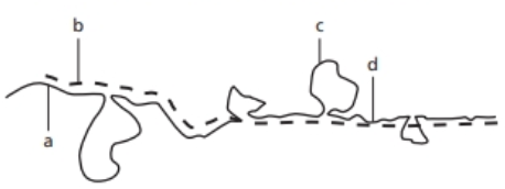
Which
Which nucleic acid is indicated by the "b" pointer? Justify your answer.
What term best identifies the nucleic acid region indicated by the "c" pointer?
What term best identifies the nucleic acid region indicated by the "d” pointer?
Based on this electron micrograph image, how many introns and exons are present in the mouse DNA fragment shown?
Want to see the full answer?
Check out a sample textbook solution
Chapter 8 Solutions
Pearson eText Genetic Analysis: An Integrated Approach -- Instant Access (Pearson+)


- Given the following eukaryotic DNA strand, transcribe and translate the DNA into apolypeptide using the 3’ – 5’ strand as the template. You may use drawings, diagrams,colours and annotations to describe how the DNA strand will be synthesized into afunctional protein. (KEY: The letters SBMD are “made up” nucleic acids that depict non-coding regions in theDNA, hypothetically S pairs with B and M pairs with D).2.2. Describe what are missense mutations and its effects on structure and function usinghaemoglobin as an example (8).5’ - TATAAAAASSMSBMDATGSBDCCMBDBAATBSMDSTGTGTCCTMSBAG – 3’arrow_forwardWe have talked about several examples of cis-acting elements that have dyad symmetry (inverted repeat symmetry). Some function on the level of DNA, and others function on the level of RNA. Give one example of one that functions at the DNA level and briefly explain why the sequence requires dyad symmetry to work properly. Note: you don't have to give an exact sequence, just the name of the element. Edit View Incort Format Tools Tabloarrow_forwardCystic fibrosis (CF) is an inherited disorder caused by different types of mutations, many of which prevent ions from moving across cell membranes. Normally there are channel proteins that allow passage of the ions, but in patients with one kind of CF these proteins seem odd. Closer examination shows that these proteins display the correct amino acid sequence. However, they fail to do their job. A) Given that the primary structure of the protein is correct, what can you infer about the DNA sequence for the gene coding this protein on this patient, is there a mutation? Explain. B) Why is the primary structure insufficient to guarantee the proper function of the protein?arrow_forward
- The sequences of several short single-stranded DNA molecules are shown below. Imagine each sequence as a typical double-stranded DNA molecule, with antiparallel strands held together by Watson-Crick base- pairs between the complementary bases. Which of these double-stranded molecules would have the highest melting temperature (Tm)? 5' ACTGAGTCTCTGACTAGTCT 3' 5' ACTTAGTCTATGACTAGTCT 3' 5' ACTTAATCTATGAATAGTCT 3' 5' ACTGCGTCTCCGACTAGTCT 3' 5' ACTGCGTCTCCGACGAGCCT 3'arrow_forwardBelow is a sequence of DNA. 5'-ttaccgataattctctctcccctcttccatgattctgattaaagaaggcgagaacgaaactatttgttaatacc-3' Using the one letter code for Amino Acids, what is the predicted AA sequence of the shortest ORF (from N to C-terminal end)? Using the one letter code for Amino Acids, what is the predicted AA sequence of the longest ORF (from N to C-terminal end)?arrow_forwardAn RNA molecule has the following percentages of bases: A = 23%, U = 42%, C = 21%, and G = 14%. Q. What would be the percentages of bases in the template strand of the DNA that contains the gene for this RNA?arrow_forward
- You have isolated a fragment of viral DNA that supposedly encodes for two proteins, 196 and 220 amino acids long respectively. The isolated DNA fragment is found to be 1100 base pairs in length. Argue why is the isolated DNA cannot code the two proteins identified earlier?arrow_forwardGiven the following Wild Type and Mutated DNA sequences: 1.) Identify where the base pair change occurs ( what letter changed?) 2.) For BOTH sequences, write the mRNA strands, define the codon regions and amino acid sequences. 3.) Describe what kind of mutation has occurred (missense, nonsense, or silent), and what effect this may have on the protein. Wild Type DNA Sequence: 3' - AGGCTCGCCTGT - 5' Mutated DNA Sequence: 3' - AGTCTCGCCTGT - 5'arrow_forwardDesign a pair of primers to amplify the entire length of the following 45 base pair sequence.Make each primer 14 bases long. Write the sequences of the primers in 5' to 3' order.(Hint: It will help for you to write out BOTH strands of the DNA sequence listed below.5'-GATGCCCGTTGGATAAATTGGGCGTCTAGAATCGGTCACACTTAG-3'arrow_forward
- Shown below is a portion of a wild-type DNA sequence that encodes the last amino acids of a protein that is 270 amino acids long. The first three bolded base pairs indicate the frame and include the coding region. 5^ ...GCTAAGTATTGCTCAAGATTAGGATGATAAATAACTGG 3^ 3^.. CGATTCATAACGAGTTCTAATCCTACTATTTATTGACC 5^ Which strand is the template strand for transcription of this gene? Briefly explain how you know. An insertion of one base pair causes the protein to decrease in length by seven amino acids. With respect to the sequence given above, where does this insertion occur? A change of one base pair leads to the protein increasing in the length by one amino acid. With respect to the sequence given above, which base pair would you change, and what would you change this base pair for the protein to increase in the length by one amino acid?arrow_forwardGiven the following eukaryotic DNA strand, transcribe and translate the DNA into a polypeptide using the 3’ – 5’ strand as the template. You may use drawings, diagrams, colours and annotations to describe how the DNA strand will be synthesized into a functional protein. (KEY: The letters SBMD are “made up” nucleic acids that depict non-coding regions in the DNA, hypothetically S pairs with B and M pairs with D).2.2. Describe what are missense mutations and its effects on structure and function using haemoglobin as an examplearrow_forwardThe E. coli DNA molecule contains 4.70 x 10⁵ base pairs. Determine the number of codons that can be present in this DNA molecule. number of codons: 1.57 x10⁶ codons Assuming that the average protein in E. coli consists of a chain of 420 amino acids, calculate the maximum number of proteins that can be coded by an E. coli DNA molecule. number of proteins: proteinsarrow_forward
 Biology: The Dynamic Science (MindTap Course List)BiologyISBN:9781305389892Author:Peter J. Russell, Paul E. Hertz, Beverly McMillanPublisher:Cengage Learning