Concept explainers
Videos
To analyze:
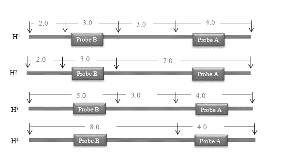
A DNA fragment is hybridized by two probes
The four resultant maps are illustrated below that show the positions, intermediate distances in “Kb” (kilobases), and the binding locations of probes with DNA. The maps correspond to alleles
In autoradiography, for genotype
In autoradiography, for genotype
If a child is born to a woman having genotype
What are expected DNA bands for each possible genotype for a child of this couple using probe A and B respectively?
Introduction:
Blotting is the technique routinely used for the detection of biomolecules such as DNA, RNA, and proteins, named as Southern blotting, northern blotting, and western blotting respectively. In southern and northern blot method, the DNA and RNA are separated on agarose gel electrophoresis and then detected by using the DNA probe complementary to the targeted sample. Southern blotting is a technique used to identify the target gene arrangement within a mixture of several genes. In
In autoradiography, the radioactively labelled substances with radioisotopes are used. In a bio-analytical procedure, such substances that are distributed in a biological sample are visualized. It is an
The first observation was recorded by Niepce de St. Victor. It was obtained accidentally when blackening occurred on the emulsion of silver chloride and Iodide by Uranium salts, and the first autoradiography used in a biological experiment traced the distribution of polonium in the specimens.
DNA preparations from different individuals normally show different patterns of the size distribution of restriction fragments that hybridize with a particular probe. These differences are called restriction fragment length polymorphisms (RFLPs). RFLP technique involves cutting a particular region of DNA with known variability, with restriction enzymes, then separating the DNA fragments by agarose gel electrophoresis, and determining the number of fragments and relative sizes.
Want to see the full answer?
Check out a sample textbook solution
Chapter 10 Solutions
GENETIC ANALYSIS: AN INTEG. APP. W/MAS


- A 2.0kb bacterial plasmid ‘BS1030’ is digested with the restriction endonuclease Sau3A; the plasmid map is depicted in the diagram below and the Sau3A (S) restriction sites are indicated. Which of the following DNA fragments do you expect to see on an agarose gel when you run Sau3A-digested plasmid ‘BS1030’ DNA? a. 250 bp, 450 bp, 550 bp, 1.1 kb, 1.5 kb and 2.0 kb b. 2.0kb c. 250 bp, 400 bp, 450 bp, 500 bp and 550 bp d. 100 bp, 200 bp, 250 bp, 400 bp, 500 bp and 550 bparrow_forwardHow would you expect your results to change if the enzyme did not have time to cut all of the sites? (Hint, remember that there are many pieces of the lambda DNA in the tube – draw yourself a simplified model where there are four restriction sites, and imagine what would happen if the enzyme only cut at one, two, three or all four, randomly, each time). How would you expect your results to change if you treated them with both EcoRI and HindIII in the same tube? Why might it be safer for the virus to have its DNA become circular when it enters the host, rather than remain linear?arrow_forwardA linear piece of DNA that is 14 kb long is cut first by EcoRI alone, then by SmaI alone, and finally, by both EcoRI and SmaI together. The following results are obtained: Draw a map of the EcoRI and SmaI restriction sites on this 14-kb piece of DNA, indicating the relative positions of the restriction sites and the distances between them.arrow_forward
- A circular plasmid of 10,000 base pairs (bp) is digested with two restriction enzymes, A and B, to produce a 3000 bp and a 2000 bp bands when visualized on an agarose gel. When digested with one enzyme at a time, only one band is visible at 5000 bp. If the first site for enzyme A (A1) is present at the 100h base, the order in which the remaining sites (A2, B1 and B2) are present is - (A) 3100, 5100, 8100 115. (B) 8100, 3100, 5100 (C) 5100, 3100, 8100 (D) 8100, 5100,. 3100arrow_forwardDNA samples from four individuals were cleaved with the same MW restriction endonuclease. The DNA fragments were separated by gel clectrophoresis, transferred to a membrane, and hybridized with a 12 kb 10 kb DNA probe complementary to a region between sites C and D (see 8 kb hybridization line). The image of the southern blot shows the labeled DNA bands and 6 kb molecular weight (MW) markers. The lane labels I, II, III, and IV -5 kb correspond to individuals I, II, III, and IV. Assume that fragments such as C-D and C-E are clearly resolved in this gel system. Fragment sizes are as given: A-B is 4 kb, B-C is I kb, C-D is 5 kb, and D-E is 650 bp. Individual I has five cleavage sites (A, B, C, D, and E) for the restriction endonuclease. DNA homologous to probe Which individual has at least one point mutation that eliminates restriction site C only? O II IV cannot be determined II Which individual has at least one point mutation that climinates restriction sites B and C? III O IV cannot be…arrow_forwardAssume that a circular plasmid is 3200 base pairs in length and has restriction sites for HindIII restriction enzyme at the following locations: 400, 700, 1400, 2600. Give the expected sizes of the restriction fragments following complete digestion.arrow_forward
- A molecule of double-stranded DNA that is 5 million base pairs long has a base composition that is 62% G + C. How many times, on average, are restriction sites for the following restriction enzymes likely to be present in this DNA molecule? a. HindIII (recognition sequence is AAGCTT)arrow_forwardIf the sequence of base pairs along a DNA molecule occurs strictly at random, what is the expected frequency of a specific restriction enzyme recognition sequence of length (a) four and (b) six base pairs?arrow_forwardWhen linear DNA is sequenced, the nucleotide base pairs are numbered from the start to finish. a DNA molecule that is 3133 base pairs long is digested with RsaI restriction enzyme recognition sites at base numbers 366, 1534, and 2207. What are the sizes of the DNA fragments that will be produced after the DNA is digested with RsaI?arrow_forward
- For the given restriction enzyme: BslI (CCNNNNN^NNGG) i) Approximately how many fragments would result from digestion of the human genome (3 × 10⁹ bases) with the enzyme? ii) Estimate the average size of the pieces of the human genome produced by digestion with the enzyme. iii) State whether the fragments of human DNA produced by digestion with the given enzyme would have sticky ends with a 5′ overhang, sticky ends with a 3′ overhang, or blunt ends. iv) If the enzyme produces sticky ends, would all the overhangs on all the ends produced by that enzyme on all fragments of the human genome be identical, or not? The recognition sequence on one strand for the enzyme is given in parentheses, with the 5′ end written at the left. N means any of the four nucleotides; R is any purine—that is, A or G; and Y is any pyrimidine—that is, C or T. ^ marks the site of cleavage. Note that the recognition sites containing Ys and Rs are not always rotationally symmetrical.arrow_forwardFour E. coli strains of genotype a+ b- are labeled 1, 2, 3, and 4. Four strains of genotype a- b+ are labeled 5, 6, 7, and 8. The two genotypes are mixed in all possible combinations and (after incubation) are plated to determine the frequency of a+ b+ recombinants. The following results are obtained, where M = many recombinants, L = low numbers of recombinants, and 0 = no recombinants:On the basis of these results, assign a sex type (either Hfr, F+, or F-) to each strain.arrow_forwardWhat is a recombinant vector? How is a recombinant vector constructed? Explain how X-Gal is used in a method of identifying recombinant vectors that contain segments of chromosomal DNA.arrow_forward
 Human Anatomy & Physiology (11th Edition)BiologyISBN:9780134580999Author:Elaine N. Marieb, Katja N. HoehnPublisher:PEARSON
 Biology 2eBiologyISBN:9781947172517Author:Matthew Douglas, Jung Choi, Mary Ann ClarkPublisher:OpenStax
 Anatomy & PhysiologyBiologyISBN:9781259398629Author:McKinley, Michael P., O'loughlin, Valerie Dean, Bidle, Theresa StouterPublisher:Mcgraw Hill Education,
 Molecular Biology of the Cell (Sixth Edition)BiologyISBN:9780815344322Author:Bruce Alberts, Alexander D. Johnson, Julian Lewis, David Morgan, Martin Raff, Keith Roberts, Peter WalterPublisher:W. W. Norton & Company
 Laboratory Manual For Human Anatomy & PhysiologyBiologyISBN:9781260159363Author:Martin, Terry R., Prentice-craver, CynthiaPublisher:McGraw-Hill Publishing Co.
 Inquiry Into Life (16th Edition)BiologyISBN:9781260231700Author:Sylvia S. Mader, Michael WindelspechtPublisher:McGraw Hill Education