Concept explainers
To analyze:
Analyzing the single amino acid substitution in the given
| Position | Amino acid | |
| Glu | ||
| Siriraj | Lys | |
| San Jose | Gly | |
| Pro | ||
| Ziguinchor | Arg | |
| Try | ||
| Bethesda | His | |
| Fort Gordon | Asp |
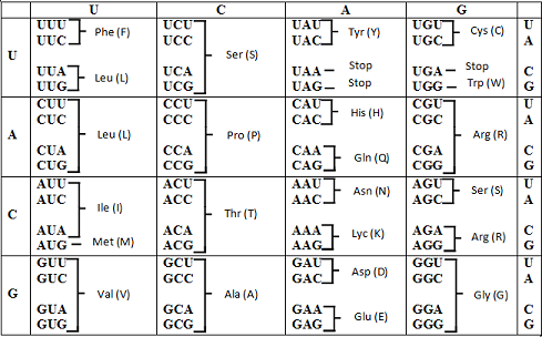
Codon table
Introduction:
The word base substitution refers to the exchange of one base pair with another. Base substitution mutation in the triplet codon can result in the synthesis of the wrong amino acid thereby changing the protein structure or function.
Many hereditary anemic conditions arise due to base substitution mutation in the wild type template strand of
Want to see the full answer?
Check out a sample textbook solution
Chapter 10 Solutions
Genetic Analysis: An Integrated Approach (2nd Edition)


- When human hemoglobin undergoes a mutation, the mutant protein usually does not replace all of the normal HbA in the red blood cells or erythrocytes of the individual. The erythro- cytes contain mixtures of varying amounts of both HbA and the mutant protein depending on the mutation and the individual. Hb Yakima is a mutant human Hb with an Asp-(B99)His mutation. The diagram on the right shows that Hb Yakima was separated by DEAE-cellulose chromatography from HbA with a 0 – 0.1 M linear gradient of NaCl buffered to pH 8.3. Why is chromatog- raphy carried out at pH 8.3? If the isoelectric point of HbA is 6.85, what is the change in total charge caused by the mutation?How does the change in charge explain the chromatography elution profile of the Hb Yakima/HbA mixture? 1,5 -Hb-A Hb -Yakima 1.0 0.5- 20 40 60 80 00 Fraction number O.D578 nmarrow_forwardYou learned in Problem 21 in Chapter 7 that theneurodegenerative disease ALS can be caused by expansion of a hexanucleotide repeat region (5′-GGGGCC-3′)outside of the open reading frame (but within the firstintron) of the gene called C9ORF72. While a normalC9ORF72 allele has 2–23 copies of the hexanucleotiderepeat unit, dominant disease-causing alleles have hundreds or even thousands of copies. Researchers observed that the first intron of theC9ORF72 disease allele is transcribed not only fromthe normal template strand of DNA, but also from thenontemplate strand. Even more unusual, both types ofrepeat-region transcripts are translated in all six readingframes in an AUG-independent manner—a processcalled repeat-associated non-ATG translation, or RANtranslation. These discoveries led to the hypothesisthat the proteins made from the repeats mightcontribute to ALS.a. What polypeptides are made from the repeat-regiontranscripts?b. According to the RAN translation hypothesis, whyare…arrow_forwardThe proximal histidine residues have been replaced by glycine residues by mutation of the cloned genes for both the α and β subunits of hemoglobin. With the tetrameric mutant hemoglobin (all subunits being mutant, α H F8 G, β H F8 G), it was found that the “proximal” coordination bonds to hemes in the mutant protein could be replaced by having the small molecule imidazole in the buffers. Oxygen binding curves for the tetrameric mutant hemoglobin were measured. A. The degree of cooperativity in oxygen binding for the mutant hemoglobin (with imidazole present) would be expected to 1) increase 2) decrease 3) not be affected) compared with the normal protein. B. Justify your answer to part A in terms of what you know about the structural basis of cooperativity in hemoglobin. C. How would the Hill coefficient for the mutant be expected to change compared with nH for normal hemoglobin, which is ~3?arrow_forward
- The protein encoded by the cystic fibrosis gene is 1480amino acids long, yet the gene spans 250 kb. How is thisdifference possible?arrow_forwardIn Sickle Cell Anemia, the production of both of the A and S beta-globin peptides in carriers suggests that the Hb beta^A and the Hb beta ^S genes^1 dominance relation is… A) Hb beta A is dominant B) Hb beta S is dominant C) Hb beta A and Hb beta S are codominant D) Hb beta A and Hb beta S are incompletely dominantarrow_forwardPeople who carry a theoretical genetic disorder (called B-disease) can be identified from a 2kb DNA sequence. People who carry this genetic disorder have a single nucleotide polymorphism that results in a change of GTATTC to GGATTC, a site that only occurs once at nucleotide number 750 in this DNA sequence. Answer the following questions based on the information provided. a) How can you develop a simple molecular test to identify the genetic disorder?arrow_forward
- A gene contains 30% thymine. What is the percentage of pyrimidines present in this segment? Explain.arrow_forwardA normal hemoglobin protein has a glutamic acid at position 6; in sickle-cell hemoglobin, this glutamic acid has been replaced by a valine. List all the possible mRNA codons that could be present for each type of hemoglobin. Can a single base change result in a change from Glu to Val in hemoglobin?arrow_forwardA gene contains the sequence CGCATACGGTAC that results in the amino acid sequence arg-ile-arq- tyr. A mutation in this gene has a G inserted after the second C in the strand. How will this mutation affect the phenotype? A)This will affect the phenotype because although most of the protein will be identical, the first amino acid will be different. B)This will not affect the phenotype because only the second amino acid is different from the original protein. C)This will not affect the phenotype because the protein will be identical to the original protein. D)This will affect the phenotype because all of the amino acids after the first one will be different from the original protein.arrow_forward
- Leber Congenital Amaurosis (LCA) causes progressive vision loss due to defects in the gene that encodes RPE65 isomerase. Affected individuals are homozygous recessive for mutant alleles of the RPE65 gene. You are trying to determine the molecular nature of the mutations in three individuals with LCA. For ease of analysis, you may assume that each individual is homozygous for the same mutant allele (though the three individuals have different mutations than each other). You use the polymerase chain reaction to amplify DNA from each patient and you determine the sequence of the DNA and compare it to unaffected individuals. You identify the following differences. Note that the non-template strand of DNA is given and the changes are highlighted using red boldface. You can assume that the sequences are in the first reading frame (eg. the first three nucleotides of each sequence is a codon). The coding region of the gene is 1602 bp and the position of the sequences shown below is…arrow_forwardHis-64 is an amino acid in myoglobin's herne-binding pocket. It is called the "distal histidine." A scientigt conducted an experiment wherein she replaced His-64 with alanine. This mutation was designated H84AA. She also created two other mutants His64AP and His64AS. She then measured the oxygen-binding ability of each of these Mb variants. Her results are given below: (a) ) What is the role of the distal Histidine in oxygen binding? (b) ) On the graph above, observe the 4 Oxygen binding curves 1, 2, 3 and 4. Curve 1 represents normal (WT) Mb. i. Variant H644A is represented by curve #: il. Variant H64AP is represented by curve # ill. Variant H64AS is represented by curve #:(c) In 3-4 sentences, explain your answers to Part (b)arrow_forwardThe genetic alteration responsible for sickle-cell anemia in humans involves: a transition mutation from A to G, substituting glutamic acid for valine in a-globin a transversion mutation from T to A, substituting valine for glutamic acid in b-globin a transition mutation from T to C, substituting valine for glutamic acid in b-globin a transversion mutation from G to C, substituting glutamic acid for valine in a-globin a frameshift mutation of one ATC codon, removing glutamic acid from b-globinarrow_forward
 Human Anatomy & Physiology (11th Edition)BiologyISBN:9780134580999Author:Elaine N. Marieb, Katja N. HoehnPublisher:PEARSON
 Biology 2eBiologyISBN:9781947172517Author:Matthew Douglas, Jung Choi, Mary Ann ClarkPublisher:OpenStax
 Anatomy & PhysiologyBiologyISBN:9781259398629Author:McKinley, Michael P., O'loughlin, Valerie Dean, Bidle, Theresa StouterPublisher:Mcgraw Hill Education,
 Molecular Biology of the Cell (Sixth Edition)BiologyISBN:9780815344322Author:Bruce Alberts, Alexander D. Johnson, Julian Lewis, David Morgan, Martin Raff, Keith Roberts, Peter WalterPublisher:W. W. Norton & Company
 Laboratory Manual For Human Anatomy & PhysiologyBiologyISBN:9781260159363Author:Martin, Terry R., Prentice-craver, CynthiaPublisher:McGraw-Hill Publishing Co.
 Inquiry Into Life (16th Edition)BiologyISBN:9781260231700Author:Sylvia S. Mader, Michael WindelspechtPublisher:McGraw Hill Education