Genetic Analysis: An Integrated Approach (2nd Edition)
2nd Edition
ISBN: 9780321948908
Author: Mark F. Sanders, John L. Bowman
Publisher: PEARSON
expand_more
expand_more
format_list_bulleted
Concept explainers
Videos
Textbook Question
Chapter 10, Problem 8P
Wild
Expert Solution & Answer
Want to see the full answer?
Check out a sample textbook solution
Students have asked these similar questions
A normal hemoglobin protein has a glutamic acid at position 6; in sickle-cell hemoglobin, this glutamic acid has been replaced by a valine. List all the possible mRNA codons that could be present for each type of hemoglobin. Can a single base change result in a change from Glu to Val in hemoglobin?
When human hemoglobin undergoes a mutation, the
mutant protein usually does not replace all of the normal HbA in
the red blood cells or erythrocytes of the individual. The erythro-
cytes contain mixtures of varying amounts of both HbA and the
mutant protein depending on the mutation and the individual. Hb
Yakima is a mutant human Hb with an Asp-(B99)His mutation.
The diagram on the right shows that Hb Yakima was separated
by DEAE-cellulose chromatography from HbA with a 0 – 0.1 M
linear gradient of NaCl buffered to pH 8.3. Why is chromatog-
raphy carried out at pH 8.3? If the isoelectric point of HbA is 6.85,
what is the change in total charge caused by the mutation?How
does the change in charge explain the chromatography elution
profile of the Hb Yakima/HbA mixture?
1,5
-Hb-A
Hb -Yakima
1.0
0.5-
20
40
60
80
00
Fraction number
O.D578 nm
Shown in the following table are several amino acid substitutionsin the a and b chains of human hemoglobin. determine how many of them can occur as a result of a single nucleotide change.
Chapter 10 Solutions
Genetic Analysis: An Integrated Approach (2nd Edition)
Ch. 10 - Define the following terms as described in this...Ch. 10 - 2. Using sickle cell disease as an example,...Ch. 10 -
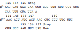
3. Compare and contrast the contributions of...Ch. 10 - Why do differences in protein electrophoretic...Ch. 10 - Prob. 5PCh. 10 - Prob. 6PCh. 10 - Prob. 7PCh. 10 - 8. Wildtype βglobin protein is composed of amino...Ch. 10 - Prob. 9PCh. 10 - Prob. 10P
Ch. 10 - 11. How is an autoradiograph produced from a...Ch. 10 - Prob. 12PCh. 10 - Prob. 13PCh. 10 - Prob. 14PCh. 10 - The family represented in the pedigree and...Ch. 10 - Suppose the mating couple (I-1 and I-2) shown in...Ch. 10 - What are restriction endonucleases, and why are...Ch. 10 - 18. Following restriction digestion, DNA fragments...Ch. 10 - 19. The doublestranded DNA sequence below is part...Ch. 10 - 20. Restriction enzymes recognize specific...Ch. 10 - Prob. 21PCh. 10 - Prob. 22PCh. 10 - Prob. 23PCh. 10 - Prob. 24PCh. 10 - 25. A second strain of dwarf plants has a...Ch. 10 - During gel electrophoresis of linear DNA...Ch. 10 - Prob. 27PCh. 10 - 28. In molecular biology, restriction...Ch. 10 - A complete plant gene containing four introns and...Ch. 10 - Prob. 30PCh. 10 - The map below illustrates three alleles in a...Ch. 10 - Prob. 32PCh. 10 - 33. Northern blot analysis is performed on mRNA...
Knowledge Booster


Learn more about
Need a deep-dive on the concept behind this application? Look no further. Learn more about this topic, biology and related others by exploring similar questions and additional content below.Similar questions
- A heptapeptide when treated with trypsin produced two peptides. T1 (D, G, Y) and T2 (K, F, V, A). When the heptapeptide was treated with chymotrypsin, three peptides were produced: CT1 (K,,Y, G), CT2 (F,A, V), and CT3 (D). The sequences of these peptides is not known, however. When the peptide was treated with Sanger’s Reagent and hydrolyzed, DNP-K and DNP-A were recovered. What is the amino acid sequence of the heptapeptide?arrow_forwardThe protein encoded by the cystic fibrosis gene is 1480amino acids long, yet the gene spans 250 kb. How is thisdifference possible?arrow_forwardIdentify the following mutations and describe what the possible effect on the protein will be. (4) 5’GAT TTT AGC TTA GCC CAT 3’ 5’ GAT TAG CTT AGC CCA T 3’ 3’CTA AAA TCG AAT CGG GTA 5’ 3’ CTA ATC GAA TCG GGT A 5’ 5’ GAT TTT AGC TTA CCC CAT 3’ 5’ GAT TTT AGC TAA CCC CAT 3’ 3’ CTA AAA TCG AAT GGG GTA 5’ 3’ CTA AAA TCG ATT GGG GTA 5’arrow_forward
- Representations of sequencing chromatograms for variants of the a chain of human hemoglobin are shown here. Match each of the variants with the corresponding amino acid change. You can use the codon table to decode each amino acid sequence. For example, the first triplet encodes for Val. Normal Chongqing ddATP ddCTP ddGTP ddTTP Pro to Thr Gly to Asp Leu to Arg Karachi Swan River Answer Bank Ala to Pro Asp to Gly Pro to Ala Arg to Leu Asp to Asn Arg to Valarrow_forwardA scientist is researching GS1, an enzyme with a relative molecular mass (Mr) of 78,000 present in a bacterium. The scientist has isolated two mutant strains of the bacterium as described below. Strain A: In this strain the GS1 protein is completely non-functional. Analysis of strain A shows that it produces a shortened GS1 protein with an Mr of only 38,000. Strain B: This produces functional GS1, but the Kcat is somewhat reduced. Analysis shows it produces a lengthened form of GS1, with an Mr of about 86,000. What type(s) of mutation may have occurred in the GS1 gene in strain A?arrow_forwardA scientist is researching GS1, an enzyme with a relative molecular mass (Mr) of 78,000 present in a bacterium. The scientist has isolated two mutant strains of the bacterium as described below. Strain A: In this strain the GS1 protein is completely non-functional. Analysis of strain A shows that it produces a shortened GS1 protein with an Mr of only 38,000. Strain B: This produces functional GS1, but the Kcat is somewhat reduced. Analysis shows it produces a lengthened form of GS1, with an Mr of about 86,000. The scientist determines the nucleotide sequence of the coding strand of the GS1 gene from strain A. It is identical to the GS1 sequence from the wild type gene except for a single change occurring approximately 1⁄3 of the way into the GS1 open reading frame. A small region of the GS1 sequence (including the site where the mutation occurs) from the wild type and mutant strains is shown below. Wild type TGTCCTCGGCCACAAGTTCTCTATC Strain A TGTCCTCGGCCACTAGTTCTCTATC How has this…arrow_forward
- A scientist is researching GS1, an enzyme with a relative molecular mass (Mr) of 78,000 present in a bacterium. The scientist has isolated two mutant strains of the bacterium as described below. Strain A: In this strain the GS1 protein is completely non-functional. Analysis of strain A shows that it produces a shortened GS1 protein with an Mr of only 38,000. Strain B: This produces functional GS1, but the Kcat is somewhat reduced. Analysis shows it produces a lengthened form of GS1, with an Mr of about 86,000. Sequencing of the GS1 gene from strain B shows that it is identical to the wild type gene except for a single alteration (the replacement of one nucleotide by another). How might this account for the features of the GS1 protein produced by strain B?arrow_forwardShown below are two homologous lengths of the alpha and betachains of human hemoglobin. Consult a genetic code dictionary and determine how many amino acid substitutionsmay have occurred as a result of a single nucleotidesubstitution. For any that cannot occur as a result of a singlechange, determine the minimal mutational distance. Alpha: ala val ala his val asp asp met proBeta: gly leu ala his leu asp asn leu lysarrow_forwardIf an extra nucleotide is inserted in the first exon of the beta globin gene, what effect will it have on the amino acid sequence of the globin polypeptides? Will the globin most likely be fully functional, partly functional, or nonfunctional? Why?arrow_forward
- Amino acid sequence analysis of all of the peptides found in a single IgG antibody would reveal unique peptide sequences totaling ~600–700 amino acids. Using this estimate, the predicted molecular weight of an antibody protein would be ~70–75 kDa. Yet, an intact antibody protein has a molecular weight of ~150 kDa. The explanation for this discrepancy is: IgG antibodies have many more heavy amino acids in them than most other proteins. Each IgG antibody is a complex of two identical light chains and two identical heavy chains. IgG antibodies tend to aggregate together during purification, thereby distorting molecular weight estimates. Each IgG antibody is a complex of four identical polypeptides. IgG antibodies are produced as dimers of two identical IgG monomers.arrow_forwardIn one type of hemoglobin variant, lysine EF6 is replaced by an aspartic acid residue. What would be the effects of this mutation on the affinity of the hemoglobin for 2,3 BPG and oxygen? The affinity for 2,3-BPG would increase, and for oxygen would decrease. The affinity for 2,3-BPG would decrease, and for oxygen would decrease. The affinities for 2,3-BPG and for oxygen would be unaffected. The affinity for 2,3-BPG would increase, and for oxygen would increase. The affinity for 2,3-BPG would decrease, and for oxygen would increase.arrow_forwardA wildtype gene produces the polypeptide sequence: Wildtype: Met-Ser-Pro-Arg-Leu-Glu-Gly Each of the following polypeptide sequences is the result of a single mutation. Identify the most likely type of mutation causing each, be as specific as possible. M1:Met-Ser-Ser-Arg-Leu-Glu-Gly missense mutation M2:Met-Ser-Pro M3:Met-Ser-Pro-Asp-Trp-Arg-Asp-Lys M4:Met-Ser-Pro-Glu-Gly nonsense mutation frameshift insertion in frame deletion M5:Met-Ser-Pro-Arg-Leu-Glu-Gly in frame insertionarrow_forward
arrow_back_ios
SEE MORE QUESTIONS
arrow_forward_ios
Recommended textbooks for you
 Human Heredity: Principles and Issues (MindTap Co...BiologyISBN:9781305251052Author:Michael CummingsPublisher:Cengage Learning
Human Heredity: Principles and Issues (MindTap Co...
Biology
ISBN:9781305251052
Author:Michael Cummings
Publisher:Cengage Learning
Biomolecules - Protein - Amino acids; Author: Tutorials Point (India) Ltd.;https://www.youtube.com/watch?v=ySNVPDHJ0ek;License: Standard YouTube License, CC-BY